WBR0948: Difference between revisions
Jump to navigation
Jump to search
No edit summary |
Sergekorjian (talk | contribs) No edit summary |
||
| Line 1: | Line 1: | ||
{{WBRQuestion | {{WBRQuestion | ||
|QuestionAuthor=William J Gibson | |QuestionAuthor=William J Gibson (Reviewed by {{YD}}) | ||
|ExamType=USMLE Step 1 | |ExamType=USMLE Step 1 | ||
|MainCategory=Physiology | |MainCategory=Physiology | ||
| Line 21: | Line 21: | ||
|MainCategory=Physiology | |MainCategory=Physiology | ||
|SubCategory=Pulmonology | |SubCategory=Pulmonology | ||
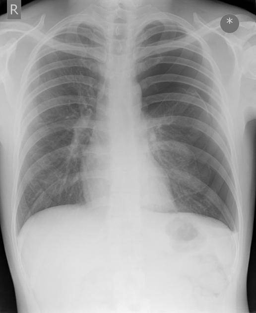
|Prompt=A 19-year-old | |Prompt=A 19-year-old man presents to the urgent care clinic with sudden onset of dyspnea and pleuritic chest pain. The patient cannot recall any recent trauma or illness. An upright chest X-ray of the patient is shown below. | ||
[[File:WBR0948.jpg | 400px]] | [[File:WBR0948.jpg | 400px]] | ||
Which of the following processes most likely explains this patient’s condition? | Which of the following processes most likely explains this patient’s condition? | ||
|Explanation=The patient in this vignette has suffered a spontanoues pneumothorax. Spontaneous pneumothorax tends to occur | |Explanation=The patient in this vignette has suffered a spontanoues pneumothorax. Spontaneous pneumothorax tends to occur among tall, thin young males (M:F ratio is 8:1). It is thought to arise from the rupture of apical "blebs" (small air-filled lesions under the pleural surface), which are presumed to be more common in those classically at risk of pneumothorax (tall males) due to mechanical factors. In spontaneous pneumothorax, blebs can be found in approximately 77% of cases, compared with only 6% in the general population without a history of spontaneous pneuomothorax. | ||
|AnswerA=Coxsackie B infection | |AnswerA=Coxsackie B infection | ||
|AnswerAExp=While Coxsackie B virus infection is the most common cause of pleurisy and pericarditis, the chest x-ray in this patient clearly | |AnswerAExp=While Coxsackie B virus infection is the most common cause of pleurisy and pericarditis, the chest x-ray in this patient clearly demonstrates a large left-sided pneumothorax. | ||
|AnswerB=Rupture of apical blebs | |AnswerB=Rupture of apical blebs | ||
|AnswerBExp=The patient in this vignette most likely has a spontaneous pneumothorax. | |AnswerBExp=The patient in this vignette most likely has a spontaneous pneumothorax, which are thought to arise from the rupture of apical blebs (small air-filled lesions under the pleural surface). | ||
|AnswerC=Fat embolism | |AnswerC=Fat embolism | ||
|AnswerCExp=While fat embolism can cause dyspnea, it is rare and usually associated with sever physical trauma such as the fracture of long bones. | |AnswerCExp=While fat embolism can cause dyspnea, it is rare and usually associated with sever physical trauma such as the fracture of long bones. | ||
|AnswerD=Deep vein thrombosis | |AnswerD=Deep vein thrombosis | ||
|AnswerDExp=The chest x-ray in this patient clearly | |AnswerDExp=The chest x-ray in this patient clearly demonstrates a large left sided pneumothorax. The patient is young and does not seem to have acquired or hereditary risk factors of deep vein thrombosis. | ||
|AnswerE=Fibrillin defect | |AnswerE=Fibrillin defect | ||
|AnswerEExp=While Marfan syndrome can cause pneumothorax, it is far more likely that this patient does not have Marfan syndrome and simply has suffered a spontaneous pneumothorax. | |AnswerEExp=While Marfan syndrome can cause pneumothorax, it is far more likely that this patient does not have Marfan syndrome and simply has suffered a spontaneous pneumothorax. | ||
|EducationalObjectives=Spontaneous pneumothorax is | |EducationalObjectives=Spontaneous pneumothorax typically occurs among tall, thin young males. It is thought to arise from the rupture of apical "blebs" (small air-filled lesions under the pleural surface). | ||
|References=Light, Richard W. "Management of spontaneous pneumothorax." | |References=Light, Richard W. "Management of spontaneous pneumothorax." Am Rev Resp Dis. 1993;148: 245-245.<br> | ||
First Aid 2015 page 615 | First Aid 2015 page 615 | ||
|RightAnswer=B | |RightAnswer=B | ||
|WBRKeyword=Pneumothorax, Lung, Pleura, Dyspnea | |WBRKeyword=Pneumothorax, Lung, Pleura, Dyspnea, Chest pain | ||
|Approved=Yes | |Approved=Yes | ||
}} | }} | ||
Revision as of 22:50, 15 August 2015
| Author | [[PageAuthor::William J Gibson (Reviewed by Yazan Daaboul, M.D.)]] |
|---|---|
| Exam Type | ExamType::USMLE Step 1 |
| Main Category | MainCategory::Physiology |
| Sub Category | SubCategory::Pulmonology |
| Prompt | [[Prompt::A 19-year-old man presents to the urgent care clinic with sudden onset of dyspnea and pleuritic chest pain. The patient cannot recall any recent trauma or illness. An upright chest X-ray of the patient is shown below.
Which of the following processes most likely explains this patient’s condition?]] |
| Answer A | AnswerA::Coxsackie B infection |
| Answer A Explanation | AnswerAExp::While Coxsackie B virus infection is the most common cause of pleurisy and pericarditis, the chest x-ray in this patient clearly demonstrates a large left-sided pneumothorax. |
| Answer B | AnswerB::Rupture of apical blebs |
| Answer B Explanation | AnswerBExp::The patient in this vignette most likely has a spontaneous pneumothorax, which are thought to arise from the rupture of apical blebs (small air-filled lesions under the pleural surface). |
| Answer C | AnswerC::Fat embolism |
| Answer C Explanation | AnswerCExp::While fat embolism can cause dyspnea, it is rare and usually associated with sever physical trauma such as the fracture of long bones. |
| Answer D | AnswerD::Deep vein thrombosis |
| Answer D Explanation | AnswerDExp::The chest x-ray in this patient clearly demonstrates a large left sided pneumothorax. The patient is young and does not seem to have acquired or hereditary risk factors of deep vein thrombosis. |
| Answer E | AnswerE::Fibrillin defect |
| Answer E Explanation | AnswerEExp::While Marfan syndrome can cause pneumothorax, it is far more likely that this patient does not have Marfan syndrome and simply has suffered a spontaneous pneumothorax. |
| Right Answer | RightAnswer::B |
| Explanation | [[Explanation::The patient in this vignette has suffered a spontanoues pneumothorax. Spontaneous pneumothorax tends to occur among tall, thin young males (M:F ratio is 8:1). It is thought to arise from the rupture of apical "blebs" (small air-filled lesions under the pleural surface), which are presumed to be more common in those classically at risk of pneumothorax (tall males) due to mechanical factors. In spontaneous pneumothorax, blebs can be found in approximately 77% of cases, compared with only 6% in the general population without a history of spontaneous pneuomothorax. Educational Objective: Spontaneous pneumothorax typically occurs among tall, thin young males. It is thought to arise from the rupture of apical "blebs" (small air-filled lesions under the pleural surface). |
| Approved | Approved::Yes |
| Keyword | WBRKeyword::Pneumothorax, WBRKeyword::Lung, WBRKeyword::Pleura, WBRKeyword::Dyspnea, WBRKeyword::Chest pain |
| Linked Question | Linked:: |
| Order in Linked Questions | LinkedOrder:: |
