WBR0901
| Author | [[PageAuthor::Serge Korjian M.D. (Reviewed by Serge Korjian)]] |
|---|---|
| Exam Type | ExamType::USMLE Step 1 |
| Main Category | MainCategory::Pathophysiology |
| Sub Category | SubCategory::Musculoskeletal/Rheumatology |
| Prompt | [[Prompt::A 62-year-old woman known to have osteoarthritis is hospitalized for pneumonia for which she receives an intravenous course of antibiotics. On her third day of hospitalization, she reports improvement in her respiratory status but complains of a new onset of knee pain that started in the morning and progressively worsened throughout the day. The knee is red, swollen and tender upon palpation. Her temperature is 37 °C (98.6 °F), blood pressure is 120/70 mmHg and heart rate of 70/min. Analysis of the synovial fluid reveals deposition of rhomboid crystals. Which other features characterizes the crystals deposited in this patient’s joint?]] |
| Answer A | AnswerA::Eosinophilic |
| Answer A Explanation | AnswerAExp::Eosinophilic crystals are characteristics of gout and not pseudogout. |
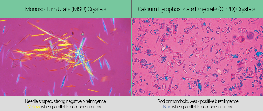
| Answer B | AnswerB::Weak positive birefringence |
| Answer B Explanation | AnswerBExp::In pseudogout, rhomboidal crystals exhibit weakly positive birefringence under polarized light. |
| Answer C | AnswerC::Yellow when parallel to light |
| Answer C Explanation | AnswerCExp::Crystals that are yellow when parallel to light are characteristic of gout and not pseudogout. |
| Answer D | AnswerD::Blue when perpendicular to light |
| Answer D Explanation | AnswerDExp::Crystals that are blue when perpendicular to light are characteristic of gout and not pseudogout. |
| Answer E | AnswerE::Hexagonal shape |
| Answer E Explanation | AnswerEExp::In pseudogout, crystals have a rhomboid shape and not a hexagonal shape. |
| Right Answer | RightAnswer::B |
| Explanation | [[Explanation::Chondrocalcinosis or pseudogout is a disorder characterized by joint inflammation due to accumulation of crystals of calcium pyrophosphate dihydrate in the connective tissues. Pseudogout resembles the gout but classically involves the larger joints very early on (eg. knee joint). There are several disorders associated with chondrocalcinosis including hemochromatosis, hyperparathyroidism, renal osteodystrophy, and hypothyroidism. Joint X-rays classically demonstrate visible calcifications in the cartilage. Synovial fluid analysis reveals rhomboidal crystals which exhibit weak positive birefringence under polarized light microscopy in contrast to the needle-shaped strongly negative birefringent crystals observed in patients with gouty arthritis.
|
| Approved | Approved::Yes |
| Keyword | WBRKeyword::Pseudogout, WBRKeyword::Crystal, WBRKeyword::Arthritis, WBRKeyword::Gout, WBRKeyword::CPPD, WBRKeyword::Chondrocalcinosis |
| Linked Question | Linked:: |
| Order in Linked Questions | LinkedOrder:: |
