WBR0775
| Author | [[PageAuthor::Serge Korjian M.D. (Reviewed by Serge Korjian)]] |
|---|---|
| Exam Type | ExamType::USMLE Step 1 |
| Main Category | MainCategory::Pathophysiology |
| Sub Category | SubCategory::Gastrointestinal, SubCategory::Musculoskeletal/Rheumatology |
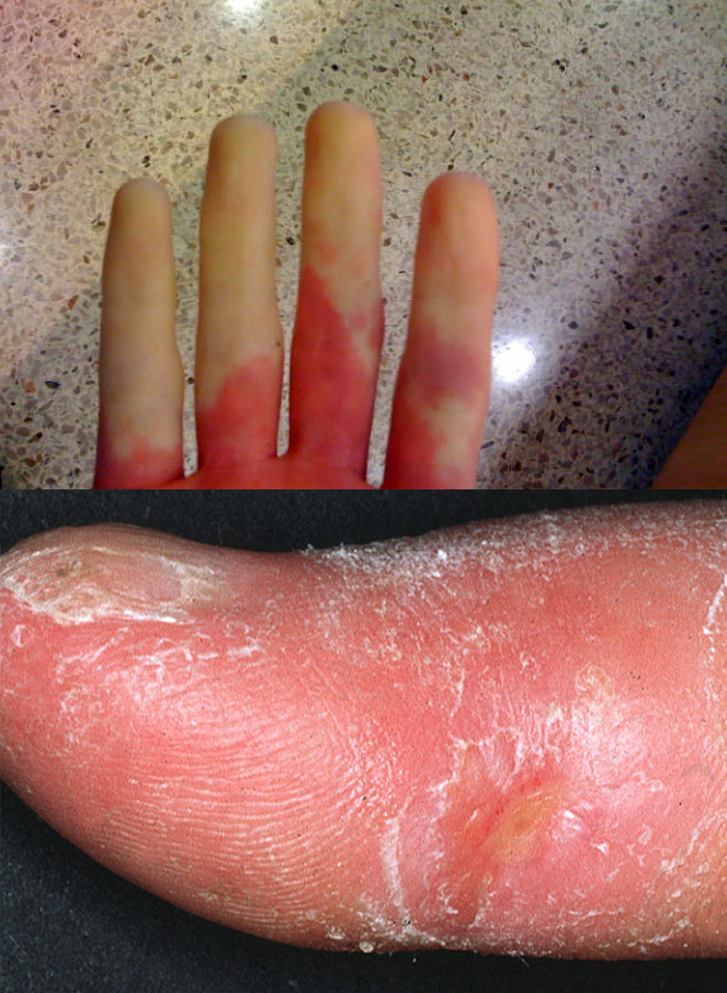
| Prompt | [[Prompt::A 48-year-old woman presents to the clinic for 3 weeks of difficulty swallowing associated with severe chest pain. The woman explains that she doesn’t remember when her symptoms started, but they have been gradually increasing. She initially had minor difficulty swallowing liquids that progressed to almost constant solid and liquid dysphagia. She has lost 5 kg (11 lbs) in the last 2 weeks because she has not been able to eat enough. Physical exam is mostly unremarkable except for the findings shown below. What antibody is most likely to be positive in this patient? |
| Answer A | AnswerA::Anti-ribonucleoprotein antibody |
| Answer A Explanation | AnswerAExp::Anti-ribonucleoprotein is usually detected in 40% of patients with SLE. |
| Answer B | AnswerB::Anti-Jo1 antibody |
| Answer B Explanation | AnswerBExp::Anti-Jo1 antibodies are found in 30-40% of patients with polymyositis and to a lesser extent in patients with dermatomyositis. |
| Answer C | AnswerC::Anticentromere antibody |
| Answer C Explanation | AnswerCExp::Anticentromere is seen in 60-70% of patients with limited scleroderma (CREST) and around 10% of patients with the diffuse form. |
| Answer D | AnswerD::Anti-double stranded DNA antibody |
| Answer D Explanation | AnswerDExp::Anti-dsDNA antibodies are very specific for SLE and are involved in the pathogenesis of lupus nephritis. |
| Answer E | AnswerE::Anti-histone antibody |
| Answer E Explanation | AnswerEExp::Anti-histone antibodies are seen in drug induced lupus. |
| Right Answer | RightAnswer::C |
| Explanation | [[Explanation::Scleroderma defines a systemic disease that is characterized by thickening and fibrosis of the skin and internal organs to varying extents depending on the subtype. Two major forms of scleroderma exist: Limited scleroderma (CREST Syndrome) and diffuse scleroderma. CREST syndrome defines a milder form referring to a clinical pentad of Calcinosis, Raynaud's phenomenon, Esophageal dysfunction, Sclerodactyly, and Telangiectasia, hence the term. The diffuse form is usually more rapidly progressive, involving large areas of skin and several internal organs. The prognosis of CREST syndrome is usually better than the diffuse form although both can be complicated by pulmonary hypertension. The etiology for both forms is unknown. Patients with CREST syndrome usually have anti-centromere antibodies while patients with diffuse scleroderma have anti-topoisomerase antibodies (anti-scl70). Treatment is mostly symptomatic. Educational Objective: CREST syndrome is the limited form of scleroderma presenting with calcinosis, Raynaud's phenomenon, esophageal dysfunction, sclerodactyly, and telangiectasia. Patients are usually is positive for anti-centromere antibody. |
| Approved | Approved::Yes |
| Keyword | WBRKeyword::CREST Syndrome, WBRKeyword::Limited Scleroderma, WBRKeyword::Systemic sclerosis, WBRKeyword::Anti-centromere antibody |
| Linked Question | Linked:: |
| Order in Linked Questions | LinkedOrder:: |
