WBR0135: Difference between revisions
Jump to navigation
Jump to search
No edit summary |
No edit summary |
||
| Line 10: | Line 10: | ||
<BR> | <BR> | ||
'''Short pathophysiology:''' increased TH1 cytokines(IL-2 and IFN-γ ) → T-cell expansion & macrophage activation, respectively → local environment: IL-8, TNF (released at high levels from alveolar macrophages → additional T cells and monocytes recruitment → non caseating granulomas → in pulmonary hilium, skin, joints and other systems. | '''Short pathophysiology:''' increased TH1 cytokines(IL-2 and IFN-γ ) → T-cell expansion & macrophage activation, respectively → local environment: IL-8, TNF (released at high levels from alveolar macrophages → additional T cells and monocytes recruitment → non caseating granulomas → in pulmonary hilium, skin, joints and other systems. | ||
Characteristically these patients present with elevated ACE (Angiotensin Converting Enzyme) levels on blood, which normally is secreted by the lungs and kidneys by cells in the inner layer of blood vessels. Its main functions are bradykinin (potent vasodilator) degradation and angiotensin II production from angiotensin I. The question asks the direct effect of [[Angiotensin-converting enzyme|ACE]]. | Characteristically these patients present with elevated [Angiotensin-converting enzyme|ACE]] (Angiotensin Converting Enzyme) levels on blood, which normally is secreted by the lungs and kidneys by cells in the inner layer of blood vessels. Its main functions are bradykinin (potent vasodilator) degradation and angiotensin II production from angiotensin I. The question asks the direct effect of [[Angiotensin-converting enzyme|ACE]]. | ||
<br> | <br> | ||
<font color="MediumBlue"><font size="4">'''Educational Objective:''' </font></font> Sarcoidosis is an autoimmune disease that causes increased serum levels of ACE, which degrades bradykinin (a potent vasodilator) and converts Angiotensin I into Angiotensin II. | <font color="MediumBlue"><font size="4">'''Educational Objective:''' </font></font> Sarcoidosis is an autoimmune disease that causes increased serum levels of [Angiotensin-converting enzyme|ACE]], which degrades bradykinin (a potent vasodilator) and converts Angiotensin I into Angiotensin II. | ||
|AnswerA=Angiotensinogen transformation | |AnswerA=Angiotensinogen transformation | ||
|AnswerAExp=<font color="red">'''Incorrect.'''</font> [[Renin]], secreted by the kidney JG cells, transforms [[Angiotensinogen]] (secreted by the liver) into Angiotensinogen I. | |AnswerAExp=<font color="red">'''Incorrect.'''</font> [[Renin]], secreted by the kidney JG cells, transforms [[Angiotensinogen]] (secreted by the liver) into Angiotensinogen I. | ||
| Line 18: | Line 18: | ||
|AnswerBExp=<font color="red">'''Incorrect.'''</font> [[Angiotensin II]] increases the ADH secretion directly. | |AnswerBExp=<font color="red">'''Incorrect.'''</font> [[Angiotensin II]] increases the ADH secretion directly. | ||
|AnswerC=Bradykinin degradation | |AnswerC=Bradykinin degradation | ||
|AnswerCExp=<font color="Green">'''Correct.'''</font> ACE degrades [[Bradykinin]], which is a potent vasodilator. | |AnswerCExp=<font color="Green">'''Correct.'''</font> [Angiotensin-converting enzyme|ACE]] degrades [[Bradykinin]], which is a potent vasodilator. | ||
|AnswerD=Angiotensin II degradation | |AnswerD=Angiotensin II degradation | ||
|AnswerDExp=<font color="red">'''Incorrect.'''</font> Angiotensin II is degraded to angiotensin III by angiotensinases located in red blood cells and the vascular beds of most tissues. It has a half-life in circulation of around 30 seconds, whereas, in tissue, it may be as long as 15–30 minutes. | |AnswerDExp=<font color="red">'''Incorrect.'''</font> Angiotensin II is degraded to angiotensin III by angiotensinases located in red blood cells and the vascular beds of most tissues. It has a half-life in circulation of around 30 seconds, whereas, in tissue, it may be as long as 15–30 minutes. | ||
Revision as of 05:18, 9 September 2013
| Author | PageAuthor::Gonzalo Romero |
|---|---|
| Exam Type | ExamType::USMLE Step 2 CK |
| Main Category | |
| Sub Category | |
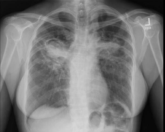
| Prompt | [[Prompt::A 50 year-old African American woman comes due to a 6-month history of dry cough. At first she thought it was an allergic reaction considering her daughter’s boyfriend recently gave a cat to her daughter. Despite intensive cleaning methods in the house, avoiding touching the cat and staying away from the cat, the cough persisted; therefore she decided to go to her primary care physician. Upon interrogation the patient states that she has been feeling fatigued during the last 4 months. She has had red and painful nodules on her shins for the last 4 weeks. Her family history is significant for autoimmune disease on her maternal side. Upon physical examination her vitals are WNL. Upon chest auscultation she has dry rales. Her lower extremities show multiple bluish-erythematous nodules, tender to palpation on her shins, varying from 3 to 5 cm in diameter. The physician orders CXR shown down below. The physician orders a blood tests in order to confirm the clinical suspicion. Which of the following is a direct function of the substance increased in this patient’s serum?
 |
| Answer A | AnswerA::Angiotensinogen transformation |
| Answer A Explanation | [[AnswerAExp::Incorrect. Renin, secreted by the kidney JG cells, transforms Angiotensinogen (secreted by the liver) into Angiotensinogen I.]] |
| Answer B | AnswerB::ADH secretion |
| Answer B Explanation | [[AnswerBExp::Incorrect. Angiotensin II increases the ADH secretion directly.]] |
| Answer C | AnswerC::Bradykinin degradation |
| Answer C Explanation | [[AnswerCExp::Correct. [Angiotensin-converting enzyme]] |
| Answer D | AnswerD::Angiotensin II degradation |
| Answer D Explanation | [[AnswerDExp::Incorrect. Angiotensin II is degraded to angiotensin III by angiotensinases located in red blood cells and the vascular beds of most tissues. It has a half-life in circulation of around 30 seconds, whereas, in tissue, it may be as long as 15–30 minutes.]] |
| Answer E | AnswerE::Angiotensin I formation |
| Answer E Explanation | [[AnswerEExp::Incorrect. Renin, secreted by the kidney JG cells, transforms Angiotensinogen (secreted from the liver) into Angiotensinogen I.]] |
| Right Answer | RightAnswer::C |
| Explanation | [[Explanation::This 50 year-old African American woman is presenting with classical clinical symptoms and signs of sarcoidosis (persistent dry cough, fatigue and bilateral hilar enlargement), which is an autoimmune chronic granulomatous disease. This patient on physical exam has erythema nodosum, which is associated with sarcoidosis. The CXR demonstrates a bilateral hiliar lymphadenopathy with minor increase in interstitial markings. She has been complaining of fatigue due to the increased levels of TNF.

|
| Approved | Approved::Yes |
| Keyword | |
| Linked Question | Linked:: |
| Order in Linked Questions | LinkedOrder:: |