Ventricular tachycardia electrocardiogram: Difference between revisions
Avirupguha (talk | contribs) (New page: == EKG Findings == 500 px|center|thumb|Ventricular tachycardia in Lead II (rhythm) # Abnormal and wide QRS complexes with se...) |
Avirupguha (talk | contribs) No edit summary |
||
| Line 1: | Line 1: | ||
[[Image:Lead II rhythm ventricular tachycardia Vtach VT.jpg|500 px|center|thumb|Ventricular tachycardia in Lead II (rhythm)]] | [[Image:Lead II rhythm ventricular tachycardia Vtach VT.jpg|500 px|center|thumb|Ventricular tachycardia in Lead II (rhythm)]] | ||
| Line 64: | Line 63: | ||
</gallery> | </gallery> | ||
</div> | </div> | ||
==References== | ==References== | ||
Revision as of 15:35, 16 September 2011

- Abnormal and wide QRS complexes with secondary ST segment and T wave changes.
- Usual QRS duration is > 0.12 seconds, may be shorter if the ectopic focus is located in the ventricular septum.
- The secondary ST segment and T wave changes are in a direction that is opposite the major deflection of the QRS.
- A ventricular rate between 140 and 200 BPM.
- When the rate is >200 and has a sine wave appearance, it is called ventricular flutter.
- When the rate is <110 BPM it is called non-paroxysmal VT.
- A regular or slightly irregular (up to 0.03 seconds) rhythm.
- Abrupt onset and termination.
- AV dissociation.
- Atrial rate slower than ventricular rate.
- No relationship between atrial activity and ventricular activity.
- There can be VA conduction.
- The RP interval is >0.11 seconds.
- Occurs in about 50% of cases.
- Uncommon when the ventricular rate is rapid (only 1/7 when the rate was>200).
- Capture beats.
- Occurs when a supraventricular impulse is conducted and captures the ventricle.
- They are rare.
- Fusion beats.
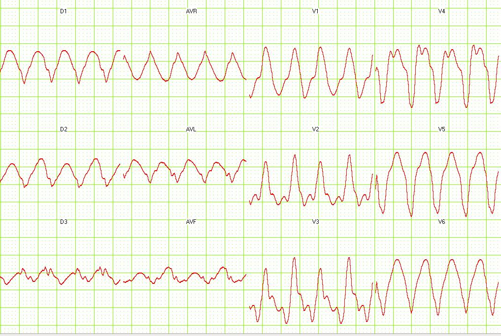
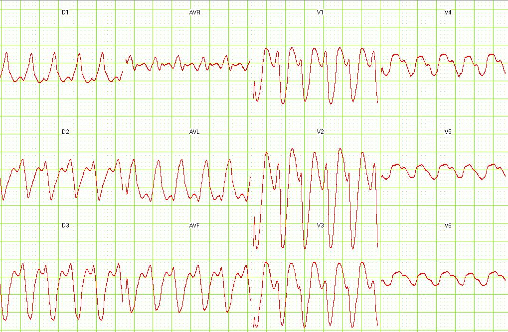
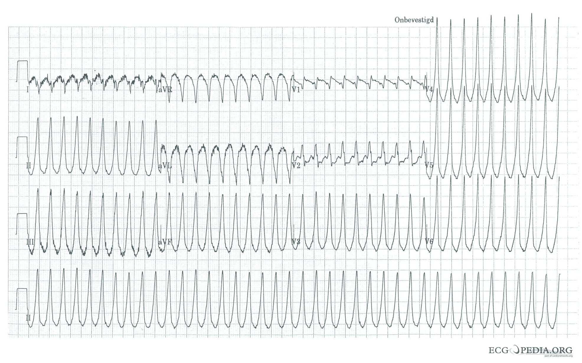
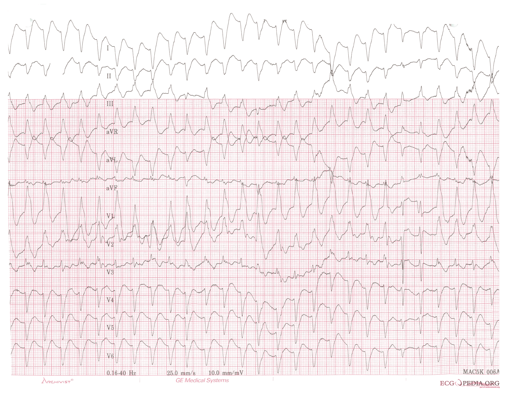
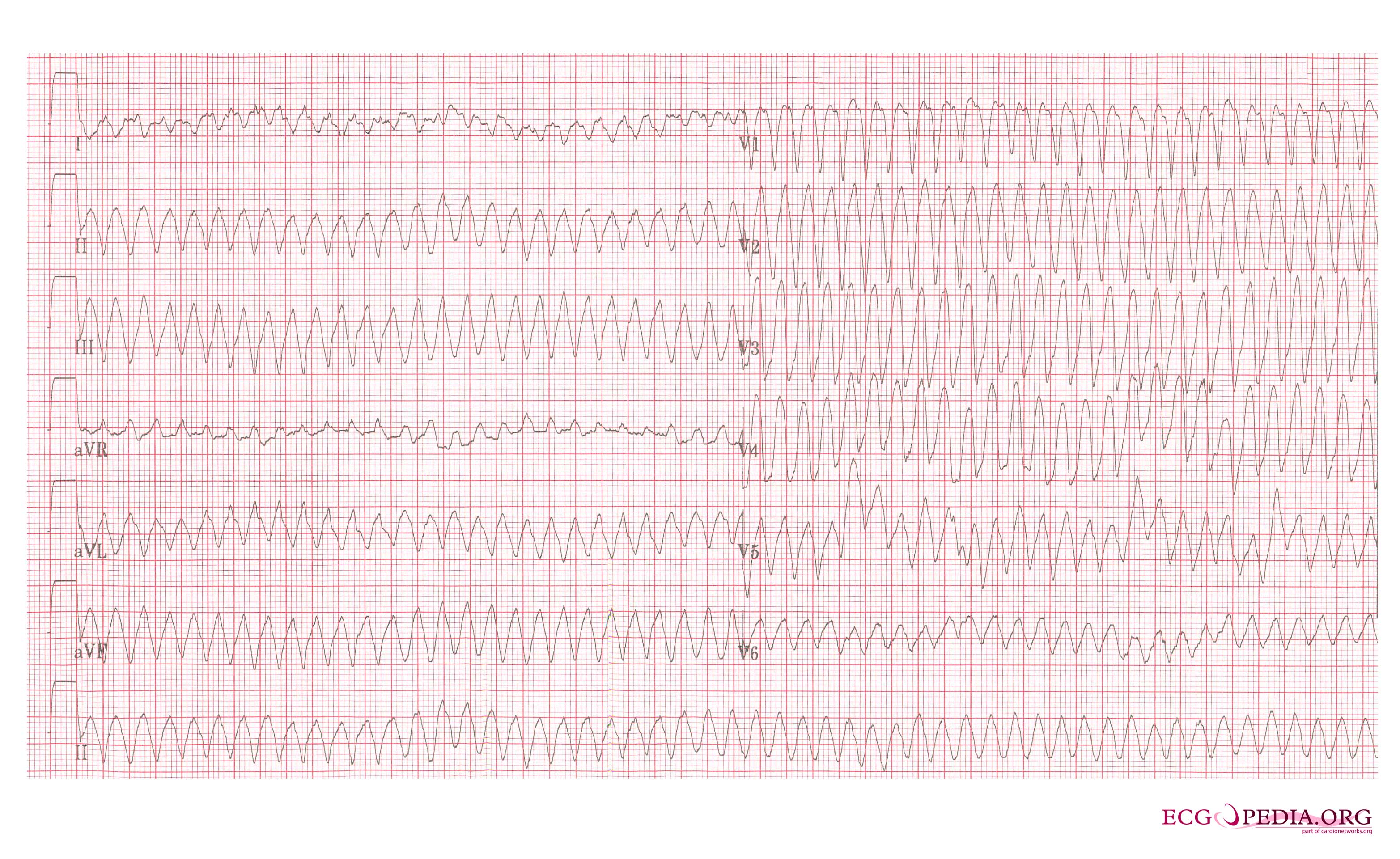
Examples of Ventricular Tachycardia:
-
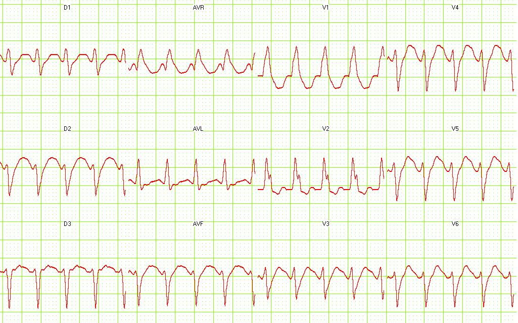
12 lead EKG: Ventricular tachycardia.
-
12 lead EKG: Ventricular tachycardia. Image courtesy of Dr Jose Ganseman


-
12 lead EKG: Ventricular tachycardia. Image courtesy of Dr Jose Ganseman
-
12 lead EKG: Ventricular tachycardia. Image courtesy of Dr Jose Ganseman


-
12 lead EKG: Ventricular tachycardia. Image courtesy of Dr Jose Ganseman
-
12 lead EKG: Ventricular tachycardia. Image courtesy of Dr Jose Ganseman


-
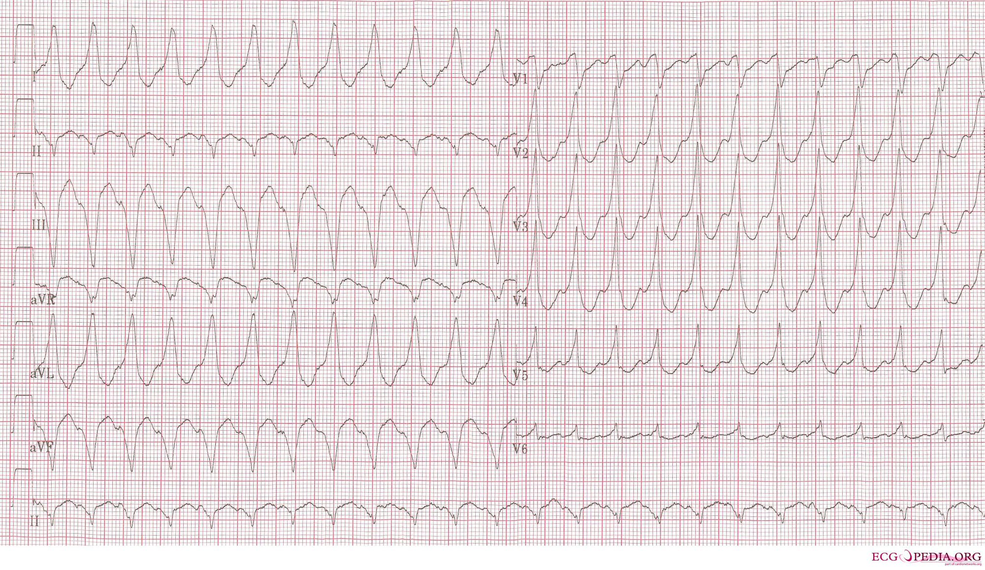
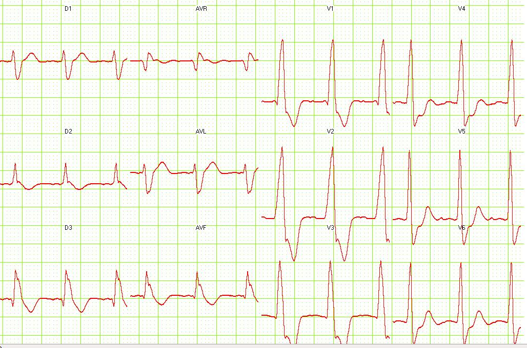
Ventricular tachycardia of 140 bpm with a left bundle branch block pattern and left heart axis.
-
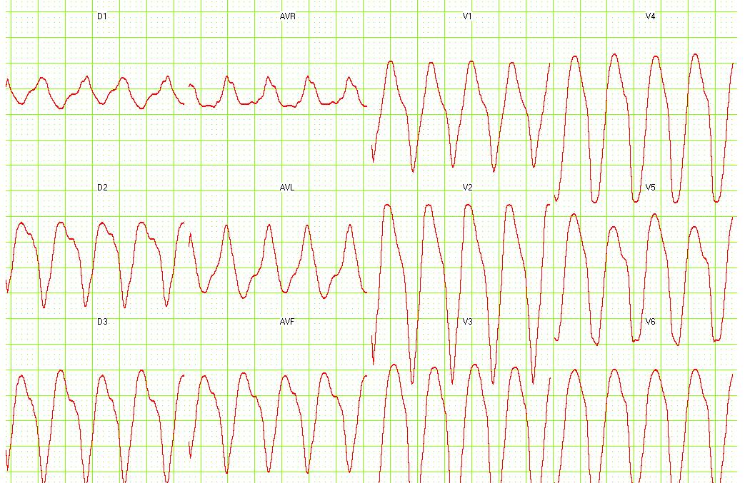
Ventricular tachycardia of 250 bpm with a right bundle branch block pattern and right heart axis.


-
Ventricular tachycardia of 150 bpm with a right bundle branch block pattern and right heart axis. Note the 5th and 6th complex from the right side. These are fusion complexes.
-
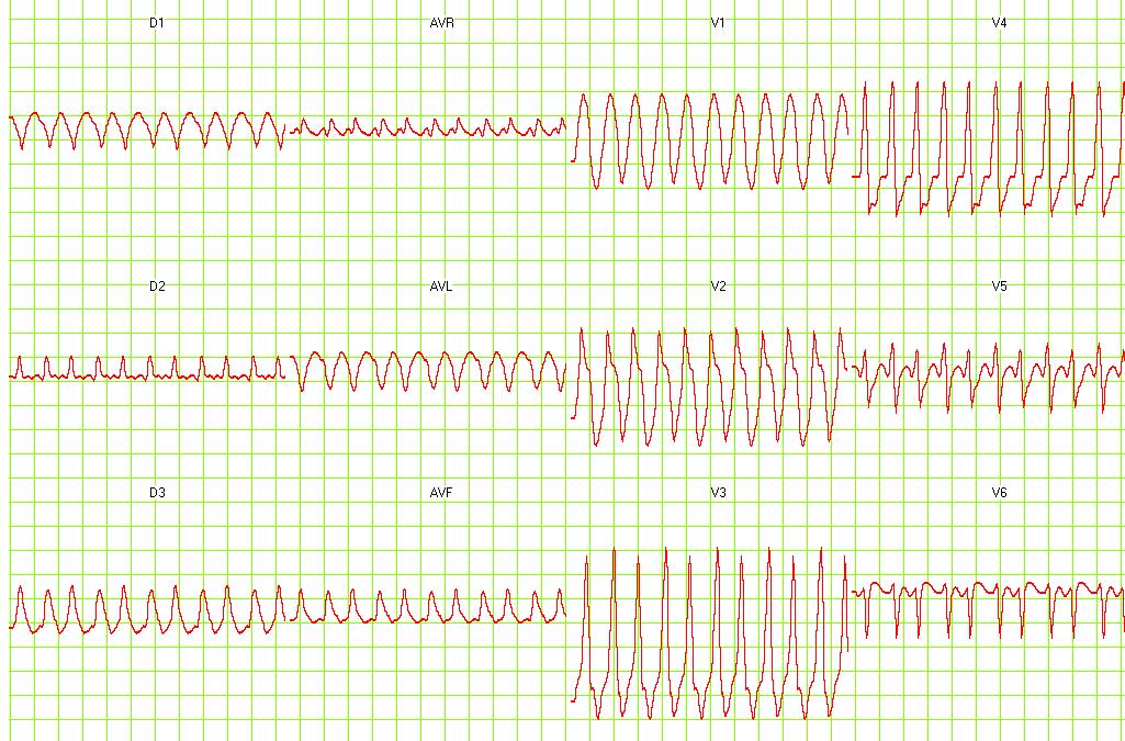
Ventricular flutter on a 12 lead ECG


References
- ↑ Chou's Electrocardiography in Clinical Practice Third Edition, pp. 398-409.
- ↑ Sailer, Christian, Wasner, Susanne. Differential Diagnosis Pocket. Hermosa Beach, CA: Borm Bruckmeir Publishing LLC, 2002:194 ISBN 1591032016
- ↑ Hammill S. C. Electrocardiographic diagnoses: Criteria and definitions of abnormalities, Chapter 18, MAYO Clinic, Concise Textbook of Cardiology, 3rd edition, 2007 ISBN 0-8493-9057-5