Ventricular fibrillation EKG examples
Editor-In-Chief: C. Michael Gibson, M.S., M.D. [1]
For the main page on ventricular fibrillation, click here
Ventricular fibrillation ECG examples
Shown below is an EKG image of ventricular fibrillation showing irregular heart rhythm, heart rate of more than 300 per minute, QRS duration unrecognizable and absent P waves.

Shown below is an EKG image of ventricular fibrillation showing irregular heart rhythm, heart rate of more than 300 per minute, QRS duration unrecognizable and absent P waves.

Shown below is an EKG image of ventricular fibrillation showing irregular heart rhythm, heart rate of more than 300 per minute, QRS duration unrecognizable and absent P waves.

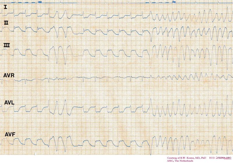
Shown below is an example of sinus rhythm converting into ventricular fibrillation wave pattern of irregular rhythm and unrecognizable QRS and P waves.

Sources
(Copyleft images obtained courtesy of ECGpedia, http://en.ecgpedia.org/index.php?title=Special:NewFiles&offset=&limit=500)