Tricuspid atresia
| Tricuspid atresia | |
 | |
|---|---|
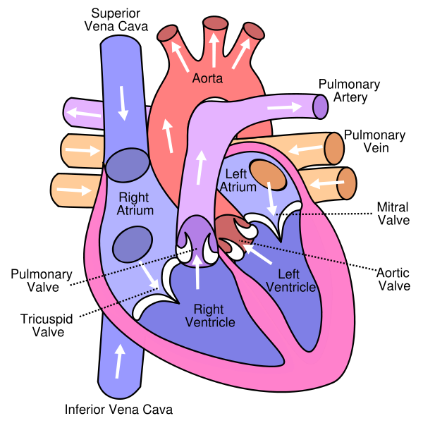
| Anterior (frontal) view of the opened heart. White arrows indicate normal blood flow. (Tricuspid valve labeled at bottom left.) | |
| ICD-10 | Q22.4 |
| ICD-9 | 746.1 |
| OMIM | 605067 |
| MedlinePlus | 001110 |
| eMedicine | med/2313 |
| MeSH | D018785 |
| Cardiology Network |
 Discuss Tricuspid atresia further in the WikiDoc Cardiology Network |
| Adult Congenital |
|---|
| Biomarkers |
| Cardiac Rehabilitation |
| Congestive Heart Failure |
| CT Angiography |
| Echocardiography |
| Electrophysiology |
| Cardiology General |
| Genetics |
| Health Economics |
| Hypertension |
| Interventional Cardiology |
| MRI |
| Nuclear Cardiology |
| Peripheral Arterial Disease |
| Prevention |
| Public Policy |
| Pulmonary Embolism |
| Stable Angina |
| Valvular Heart Disease |
| Vascular Medicine |
Editor-In-Chief: C. Michael Gibson, M.S., M.D. [1]
Associate Editor-in-Chief: Keri Shafer, M.D. [2]
Please Join in Editing This Page and Apply to be an Editor-In-Chief for this topic: There can be one or more than one Editor-In-Chief. You may also apply to be an Associate Editor-In-Chief of one of the subtopics below. Please mail us [3] to indicate your interest in serving either as an Editor-In-Chief of the entire topic or as an Associate Editor-In-Chief for a subtopic. Please be sure to attach your CV and or biographical sketch.
Overview
Tricuspid atresia is a form of congenital heart disease whereby there is a complete absence of the tricuspid valve. Therefore, there is an absence of right atrioventricular connection. This leads to a hypoplastic or an absence of the right ventricle.
Pathophysiology
This defect occurs during prenatal development. Because of the lack of an A-V connection, an atrial septal defect (ASD) must be present to maintain blood flow. Also, since there is a lack of a right ventricle there must be a way to pump blood into the pulmonary arteries, and this is accomplished by a ventricular septal defect (VSD).
Blood is mixed in the left atrium. Because the only way the pulmonary circulation receives blood is through the VSD, a patent ductus arteriosus is usually also formed to increase pulmonary flow.
Clinical manifestations
- progressive cyanosis
- Poor feeding
- Tachypnea over the first 2 weeks of life
- Holosystolic murmur due to the VSD
- Superior axis deviation and left ventricular hypertrophy (since it must pump blood to both the pulmonary and systemic systems)
- normal heart size
Treatment
- PGE1 to maintain patent ductus arteriosus
- Modified Blalock-Taussig shunt to maintain pulmonary blood flow by placing a Gortex conduit between the subclavian artery and the pulmonary artery.
- Cavopulmonary anastomosis (hemi-Fontan or bidirectional Glenn) to provide stable pulmonary flow
- Fontan procedure to redirect inferior vena cava and hepatic vein flow into the pulmonary circulation
External Links
- Tricuspid Atresia information from Seattle Children's Hospital Heart Center
Additional Reading
- Moss and Adams' Heart Disease in Infants, Children, and Adolescents Hugh D. Allen, Arthur J. Moss, David J. Driscoll, Forrest H. Adams, Timothy F. Feltes, Robert E. Shaddy, 2007 ISBN 0781786843