Torsade de pointes: Difference between revisions
Jump to navigation
Jump to search
| Line 38: | Line 38: | ||
* [[Cardiac enzymes]] | * [[Cardiac enzymes]] | ||
* [[Echocardiography]] to rule out structural heart disease | * [[Echocardiography]] to rule out structural heart disease | ||
==Additional Information== | ==Additional Information== | ||
Revision as of 01:54, 15 October 2012
| Torsade de pointes | |
 | |
|---|---|
| DiseasesDB | 29252 |
| MeSH | D016171 |
|
Torsades de pointes Microchapters |
|
Diagnosis |
|---|
|
Treatment |
|
Case Studies |
|
Torsade de pointes On the Web |
|
American Roentgen Ray Society Images of Torsade de pointes |
Editor-In-Chief: C. Michael Gibson, M.S., M.D. [1]; Associate Editor(s)-In-Chief: Cafer Zorkun, M.D., Ph.D. [2]
Clinical Correlation
- Drugs: quinidine, PCA, norpace, amiodarone, phenothiazines, Tricyclic antidepressants, pentamidine.
- with quinidine majority of the cases occur within one week of initiation, and with therapeutic levels
- Electrolyte imbalances: Hypokalemia, hypomagnesemia, hypocalcemia
- CAD
- MVP
- Variant angina
- Myocarditis
- Subarachnoid hemorrhage
- Congenital QT prolongation
- Liquid protein diets
- Hypothyroidism
- because of bradycardia and a prolonged QT syndrome
- Organophosphate poisoning [1] [2]
Other lab studies
- Electrolytes levels to rule out hypokalemia, hypomagnesemia, and hypocalcemia.
- Cardiac enzymes
- Echocardiography to rule out structural heart disease
Additional Information
Examples
EKG's shown below are courtesy of C. Michael Gibson MS MD, and copylefted
-
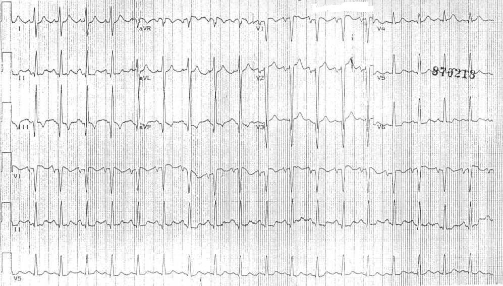
12 lead EKG at admission
-
-
-
-
-
-
-
-
-
-
-


Examples from different resources
-
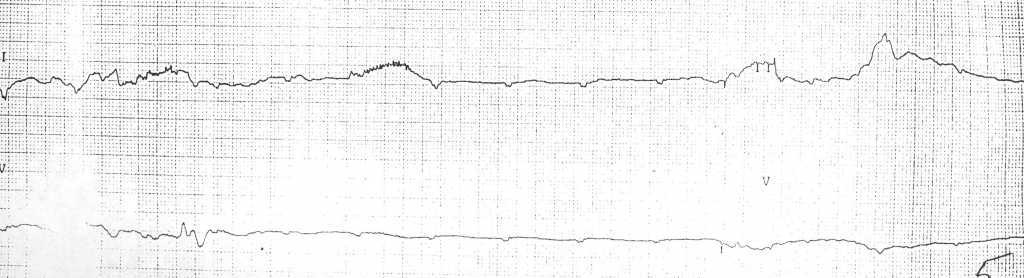
![Arrhythmias in a patient with short coupled torsade de pointes[3]](/images/f/f1/Shortcoupled_tdp1.jpg)
Arrhythmias in a patient with short coupled torsade de pointes[3]
-
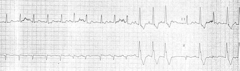
![Arrhythmias in a patient with short coupled torsades de pointes degenerating in ventricular fibrillation[3]](/images/2/2c/Shortcoupled_tdp2.jpg)
Arrhythmias in a patient with short coupled torsades de pointes degenerating in ventricular fibrillation[3]
![Arrhythmias in a patient with short coupled torsade de pointes[3]](/index.php/File:Shortcoupled_tdp1.jpg)
![Arrhythmias in a patient with short coupled torsades de pointes degenerating in ventricular fibrillation[3]](/index.php/File:Shortcoupled_tdp2.jpg)
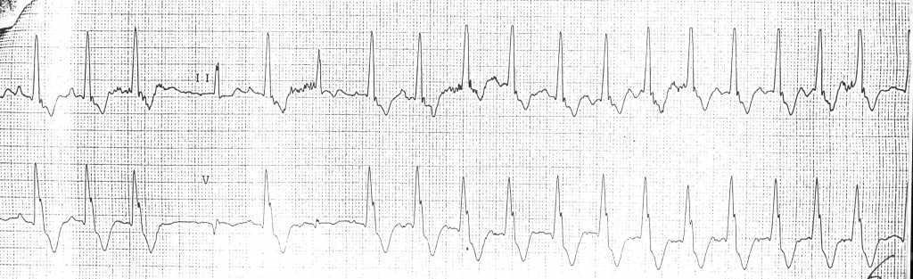
![Arrhythmias in a patient with short coupled torsade de pointes: frequent short coupled extrasystoles[3]](/index.php/File:Shortcoupled_tdp3.jpg)
![Arrhythmias in a patient with short coupled torsade de pointes: frequent short coupled extrasystoles [3]](/index.php/File:Shortcoupled_tdp4.jpg)
-
![A 12 lead ECG recording example of TdP[4]](/images/f/fc/12leadTorsade.jpg)
A 12 lead ECG recording example of TdP[4]
![A 12 lead ECG recording example of TdP[4]](/index.php/File:12leadTorsade.jpg)
References
- ↑ Chou's Electrocardiography in Clinical Practice Third Edition, pp. 398-409.
- ↑ Sailer, Christian, Wasner, Susanne. Differential Diagnosis Pocket. Hermosa Beach, CA: Borm Bruckmeir Publishing LLC, 2002:194 ISBN 1591032016
- ↑ 3.0 3.1 3.2 3.3 Leenhardt A, Glaser E, Burguera M, Nuernberg M, Maison-Blanche P, and Coumel P. Short-coupled variant of torsade de pointes. A new electrocardiographic entity in the spectrum of idiopathic ventricular tachyarrhythmias. Circulation 1994 Jan; 89(1) 206-15. PMID 8281648
- ↑ Khan IA. Twelve-lead electrocardiogram of torsade de pointes Tex Heart Inst J. 2001; 28 (1): 69. PMID 11330748