Systemic lupus erythematosus ultrasound or echocardiography: Difference between revisions
No edit summary |
|||
| (4 intermediate revisions by 2 users not shown) | |||
| Line 1: | Line 1: | ||
__NOTOC__ | __NOTOC__ | ||
{{Systemic lupus erythematosus}} | {{Systemic lupus erythematosus}} | ||
{{CMG}} {{AE}}{{MIR}} | {{CMG}}; {{AE}} {{MIR}} | ||
==Overview== | ==Overview== | ||
On abdominal [[ultrasound]], systemic lupus erythematosus (SLE) may present with [[hepatosplenomegaly]], [[ascites]], hyperecho-kidney tissue due to [[nephritis]], and rarely [[cholecystitis]]. On synovial [[ultrasound]], SLE may present with synovial effusions and [[synovitis]]. On echocardiography, SLE may present with decreased [[ejection fraction]], cardiac wall motion abnormality, [[Pericarditis|effusion pericarditis]], and valve leaflet thickening. | On abdominal [[ultrasound]], systemic lupus erythematosus (SLE) may present with [[hepatosplenomegaly]], [[ascites]], hyperecho-kidney tissue due to [[nephritis]], and rarely [[cholecystitis]]. On synovial [[ultrasound]], SLE may present with synovial effusions and [[synovitis]]. On echocardiography, SLE may present with decreased [[ejection fraction]], cardiac wall motion abnormality, [[Pericarditis|effusion pericarditis]], and valve leaflet thickening. | ||
| Line 31: | Line 31: | ||
**[[Gallbladder]] distension | **[[Gallbladder]] distension | ||
| | | | ||
[[File:3fc11253ba09067fb09f32399ba387 big gallery.jpg|thumb|300px|<SMALL><SMALL>''[https://radiopaedia.org/ | [[File:3fc11253ba09067fb09f32399ba387 big gallery.jpg|thumb|300px|<SMALL><SMALL>''[https://radiopaedia.org/ Adapted from Radiopaedia]''</SMALL></SMALL>]] | ||
[[File:Acute-acalculous-cholecystitis-1.jpg|thumb|300px|<SMALL><SMALL>''[https://radiopaedia.org/ | [[File:Acute-acalculous-cholecystitis-1.jpg|thumb|300px|<SMALL><SMALL>''[https://radiopaedia.org/ Adapted from Radiopaedia]''</SMALL></SMALL>]] | ||
|- | |- | ||
| style="background: #DCDCDC; " |<small><small>[[Renal]]</small></small> | | style="background: #DCDCDC; " |<small><small>[[Renal]]</small></small> | ||
| Line 48: | Line 48: | ||
** Echo-free space between the [[Visceral pleura|visceral]] and [[parietal pleura]] | ** Echo-free space between the [[Visceral pleura|visceral]] and [[parietal pleura]] | ||
| | | | ||
[[File:Subpulmonic effusion on ultrasonography.jpg|thumb|300px|<SMALL><SMALL>''[https://radiopaedia.org/ | [[File:Subpulmonic effusion on ultrasonography.jpg|thumb|300px|<SMALL><SMALL>''[https://radiopaedia.org/ Adapted from Radiopaedia]''</SMALL></SMALL>]] | ||
|- | |- | ||
| style="background: #DCDCDC; " |<small><small>[[Joints]]</small></small> | | style="background: #DCDCDC; " |<small><small>[[Joints]]</small></small> | ||
| Line 58: | Line 58: | ||
** Global thickening with effusion in the sheath of [[tendon]] | ** Global thickening with effusion in the sheath of [[tendon]] | ||
| | | | ||
[[File:Extensor-carpi-ulnaris-tenosynovitis-1.jpg|thumb|300px|<SMALL><SMALL>''[https://radiopaedia.org/ | [[File:Extensor-carpi-ulnaris-tenosynovitis-1.jpg|thumb|300px|<SMALL><SMALL>''[https://radiopaedia.org/ Adapted from Radiopaedia]''</SMALL></SMALL>]] | ||
|- | |- | ||
| style="background: #DCDCDC; " |<small><small>[[Raynaud phenomenon]]</small></small> | | style="background: #DCDCDC; " |<small><small>[[Raynaud phenomenon]]</small></small> | ||
| Line 68: | Line 68: | ||
== Echocardiography == | == Echocardiography == | ||
{| style="border: 3px; font-size: 100%; " | |||
| style="background:#FFFFFF;" |Main [[Echocardiography|echocardiographic]] findings in SLE include:<ref name="pmid2372888">{{cite journal |vauthors=Nihoyannopoulos P, Gomez PM, Joshi J, Loizou S, Walport MJ, Oakley CM |title=Cardiac abnormalities in systemic lupus erythematosus. Association with raised anticardiolipin antibodies |journal=Circulation |volume=82 |issue=2 |pages=369–75 |year=1990 |pmid=2372888 |doi= |url=}}</ref><ref name="pmid24599923">{{cite journal |vauthors=Hübbe-Tena C, Gallegos-Nava S, Márquez-Velasco R, Castillo-Martínez D, Vargas-Barrón J, Sandoval J, Amezcua-Guerra LM |title=Pulmonary hypertension in systemic lupus erythematosus: echocardiography-based definitions predict 6-year survival |journal=Rheumatology (Oxford) |volume=53 |issue=7 |pages=1256–63 |year=2014 |pmid=24599923 |doi=10.1093/rheumatology/keu012 |url=}}</ref> | |||
* Decreased [[ejection fraction]] | |||
* | |||
* [[Myocarditis]] | * [[Myocarditis]] | ||
** Wall motion abnormality diagnosed mostly by trans-esophageal [[echocardiography]] | ** Wall motion abnormality diagnosed mostly by trans-esophageal [[echocardiography]] | ||
| Line 80: | Line 79: | ||
* [[Pericardial effusion]] | * [[Pericardial effusion]] | ||
** [[Echocardiography]] is the method of choice to confirm the diagnosis, estimate the volume of fluid and most importantly assess the haemodynamic impact of the effusion | ** [[Echocardiography]] is the method of choice to confirm the diagnosis, estimate the volume of fluid and most importantly assess the haemodynamic impact of the effusion | ||
|<br><br>[[File:5e2515ac54c842fffa820c85e60acd big gallery.jpeg|thumb|right|500px|<SMALL><SMALL>''[https://radiopaedia.org/ Adapted from Radiopaedia]''</SMALL></SMALL>]] | |||
|} | |||
==Refrences== | ==Refrences== | ||
{{reflist|2}} | {{reflist|2}} | ||
Latest revision as of 12:03, 17 August 2017
|
Systemic lupus erythematosus Microchapters |
|
Differentiating Systemic lupus erythematosus from other Diseases |
|---|
|
Diagnosis |
|
Treatment |
|
Case Studies |
|
Systemic lupus erythematosus ultrasound or echocardiography On the Web |
|
American Roentgen Ray Society Images of Systemic lupus erythematosus ultrasound or echocardiography |
|
FDA on Systemic lupus erythematosus ultrasound or echocardiography |
|
on Systemic lupus erythematosus ultrasound or echocardiography |
|
Systemic lupus erythematosus ultrasound or echocardiography in the news |
|
Blogs onSystemic lupus erythematosus ultrasound or echocardiography |
|
Directions to Hospitals Treating Systemic lupus erythematosus |
|
Risk calculators and risk factors for Systemic lupus erythematosus ultrasound or echocardiography |
Editor-In-Chief: C. Michael Gibson, M.S., M.D. [1]; Associate Editor(s)-in-Chief: Mahshid Mir, M.D. [2]
Overview
On abdominal ultrasound, systemic lupus erythematosus (SLE) may present with hepatosplenomegaly, ascites, hyperecho-kidney tissue due to nephritis, and rarely cholecystitis. On synovial ultrasound, SLE may present with synovial effusions and synovitis. On echocardiography, SLE may present with decreased ejection fraction, cardiac wall motion abnormality, effusion pericarditis, and valve leaflet thickening.
Ultrasound
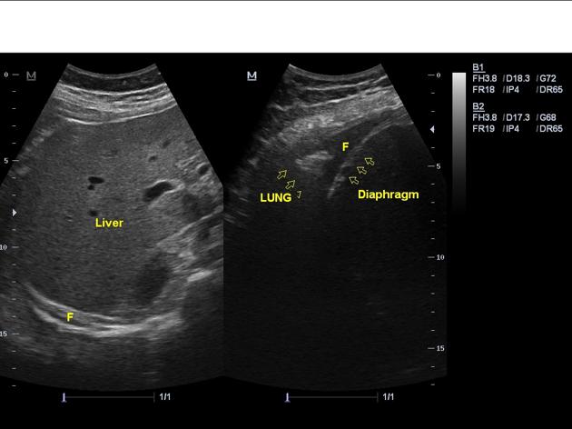
Ultrasound can be used for the diagnosis of systemic lupus erythematosus complications. It can also be used for screening and monitoring the disease activity during pregnancy.[1] The table below presents the main ultrasound findings regarding the organ system involvement in SLE:[2][3][4][5]
| Organ | Sonography findings | Preview |
|---|---|---|
| Gastrointestinal |
|
  |
| Renal |
|
|
| Pulmonary |
|
 |
| Joints |
 | |
| Raynaud phenomenon |
|
Echocardiography
Main echocardiographic findings in SLE include:[6][7]
|
 |
Refrences
- ↑ Giancotti A, Spagnuolo A, Bisogni F, D'Ambrosio V, Pasquali G, Panici PB (2011). "Pregnancy and systemic lupus erythematosus: role of ultrasound monitoring". Eur. J. Obstet. Gynecol. Reprod. Biol. 154 (2): 233–4. doi:10.1016/j.ejogrb.2010.10.020. PMID 21144639.
- ↑ Lins CF, Santiago MB (2015). "Ultrasound evaluation of joints in systemic lupus erythematosus: a systematic review". Eur Radiol. 25 (9): 2688–92. doi:10.1007/s00330-015-3670-y. PMID 25716942.
- ↑ Virdi RP, Bashir A, Shahzad G, Iqbal J, Mejia JO (2012). "Diffuse alveolar hemorrhage: a rare life-threatening condition in systemic lupus erythematosus". Case Rep Pulmonol. 2012: 836017. doi:10.1155/2012/836017. PMC 3420594. PMID 22934226.
- ↑ Ossandon A, Iagnocco A, Alessandri C, Priori R, Conti F, Valesini G (2009). "Ultrasonographic depiction of knee joint alterations in systemic lupus erythematosus". Clin. Exp. Rheumatol. 27 (2): 329–32. PMID 19473577.
- ↑ Iagnocco A, Ceccarelli F, Rizzo C, Truglia S, Massaro L, Spinelli FR, Vavala C, Valesini G, Conti F (2014). "Ultrasound evaluation of hand, wrist and foot joint synovitis in systemic lupus erythematosus". Rheumatology (Oxford). 53 (3): 465–72. doi:10.1093/rheumatology/ket376. PMID 24231444.
- ↑ Nihoyannopoulos P, Gomez PM, Joshi J, Loizou S, Walport MJ, Oakley CM (1990). "Cardiac abnormalities in systemic lupus erythematosus. Association with raised anticardiolipin antibodies". Circulation. 82 (2): 369–75. PMID 2372888.
- ↑ Hübbe-Tena C, Gallegos-Nava S, Márquez-Velasco R, Castillo-Martínez D, Vargas-Barrón J, Sandoval J, Amezcua-Guerra LM (2014). "Pulmonary hypertension in systemic lupus erythematosus: echocardiography-based definitions predict 6-year survival". Rheumatology (Oxford). 53 (7): 1256–63. doi:10.1093/rheumatology/keu012. PMID 24599923.