Suprarenal veins: Difference between revisions
Jump to navigation
Jump to search
m (Robot: Automated text replacement (-{{SIB}} +, -{{EH}} +, -{{EJ}} +, -{{Editor Help}} +, -{{Editor Join}} +)) |
m (Robot: Automated text replacement (-{{reflist}} +{{reflist|2}}, -<references /> +{{reflist|2}}, -{{WikiDoc Cardiology Network Infobox}} +)) |
||
| Line 14: | Line 14: | ||
MeshNumber = | | MeshNumber = | | ||
}} | }} | ||
{{CMG}} | {{CMG}} | ||
Latest revision as of 15:11, 6 September 2012
Editor-In-Chief: C. Michael Gibson, M.S., M.D. [1]
Overview
The Suprarenal Veins are two in number:
- the right ends in the inferior vena cava.
- the left ends in the left renal or left inferior phrenic vein.
They receive blood from the adrenal glands and will sometimes form anastomoses with the inferior phrenic veins.
Additional images
-
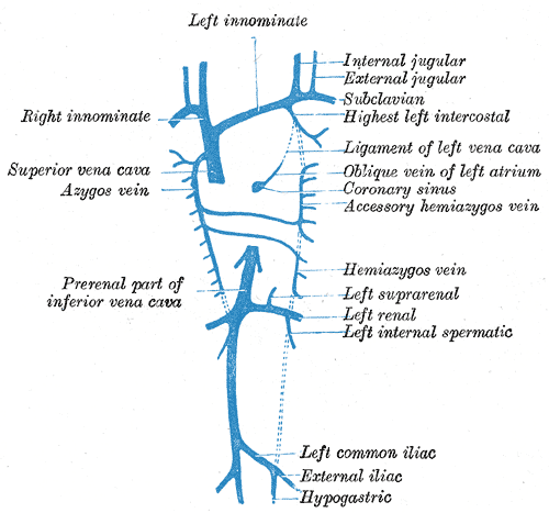
Diagram showing completion of development of the parietal veins.
-
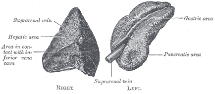
Suprarenal glands viewed from the front.
-
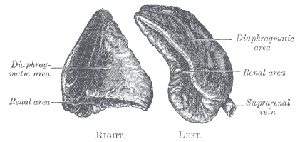
Suprarenal glands viewed from behind.


External links
- Template:SUNYAnatomyLabs - "Posterior Abdominal Wall: Blood Supply to the Suprarenal Glands"
- Template:Dorlands - left
- Template:Dorlands - right