Superior rectal artery
| Cardiology Network |
 Discuss Superior rectal artery further in the WikiDoc Cardiology Network |
| Adult Congenital |
|---|
| Biomarkers |
| Cardiac Rehabilitation |
| Congestive Heart Failure |
| CT Angiography |
| Echocardiography |
| Electrophysiology |
| Cardiology General |
| Genetics |
| Health Economics |
| Hypertension |
| Interventional Cardiology |
| MRI |
| Nuclear Cardiology |
| Peripheral Arterial Disease |
| Prevention |
| Public Policy |
| Pulmonary Embolism |
| Stable Angina |
| Valvular Heart Disease |
| Vascular Medicine |
Editor-In-Chief: C. Michael Gibson, M.S., M.D. [1]
The superior rectal artery (superior hemorrhoidal artery) is an artery that descends into the pelvis to supply blood to the rectum.
Structure
The superior rectal artery is the continuation of the inferior mesenteric artery. It descends into the pelvis between the layers of the mesentery of the sigmoid colon, crossing the left common iliac artery and vein.
It divides, opposite the third sacral vertebra into two branches, which descend one on either side of the rectum. About 10 or 12 cm from the anus, these branches break up into several small branches.
These pierce the muscular coat of the bowel and run downward, as straight vessels, placed at regular intervals from each other in the wall of the gut between its muscular and mucous coats, to the level of the internal anal sphincter; here they form a series of loops around the lower end of the rectum, and communicate with the middle rectal artery (from the internal iliac artery) and with the inferior rectal artery (from the internal pudendal artery).
Additional images
-
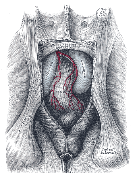
The posterior aspect of the rectum exposed by removing the lower part of the sacrum and the coccyx.

See also
External links
- Template:SUNYAnatomyFigs - "Branches of the inferior mesenteric artery."
- Template:SUNYAnatomyLabs - "Intestines and Pancreas: Branches of the Inferior Mesenteric Artery"
- Template:NormanAnatomy
- Template:NormanAnatomy (Template:NormanAnatomyFig)