Uploads by Mmir
Jump to navigation
Jump to search
This special page shows all uploaded files.
| Date | Name | Thumbnail | Size | Description | Versions |
|---|---|---|---|---|---|
| 17:45, 13 June 2017 | Molluscumcontagiosum1.jpg (file) |  |
27 KB | 1 | |
| 23:36, 13 June 2017 | Molluscaklein.jpg (file) |  |
142 KB | 2 | |
| 23:10, 28 July 2017 | Lupus final.jpg (file) |  |
154 KB | 1 | |
| 21:55, 31 July 2017 | Vacuolar interface dermatitis - high mag.jpg (file) |  |
388 KB | 1 | |
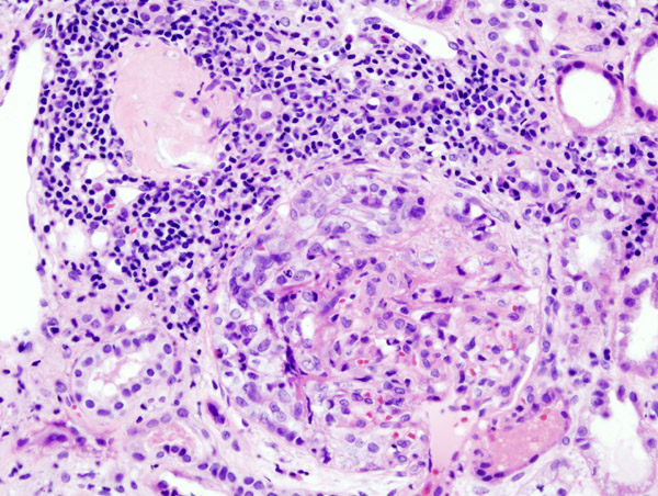
| 22:55, 31 July 2017 | Crescentic glomerulonephritis (2).jpg (file) | .jpg) |
160 KB | 1 | |
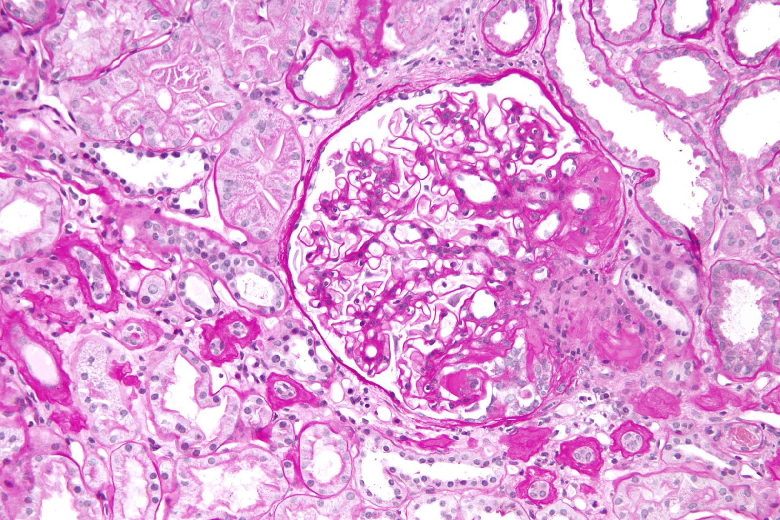
| 22:56, 31 July 2017 | 1599px-Focal segmental glomerulosclerosis - high mag.jpg (file) |  |
389 KB | 1 | |
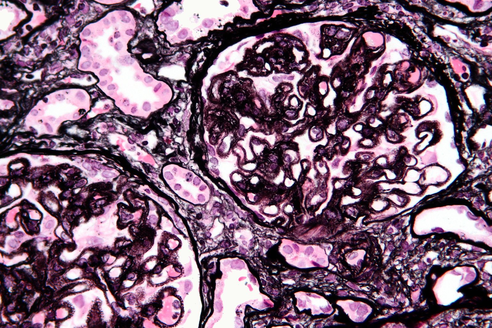
| 22:57, 31 July 2017 | Membranous nephropathy - mpas - very high mag.jpg (file) |  |
406 KB | 1 | |
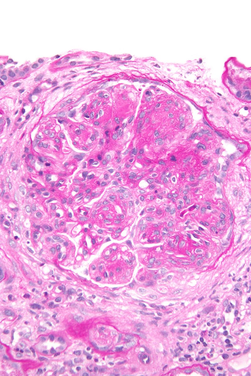
| 22:59, 31 July 2017 | Membranoproliferative glomerulonephritis - very high mag.jpg (file) |  |
192 KB | 1 | |
| 00:33, 1 August 2017 | Lupus2.jpg (file) |  |
154 KB | 1 | |
| 00:38, 1 August 2017 | Thumbprinting xray.jpeg (file) |  |
132 KB | 1 | |
| 00:39, 1 August 2017 | Thumbprinting ct.jpeg (file) |  |
65 KB | 1 | |
| 00:44, 1 August 2017 | Pleural effusion ct.jpg (file) |  |
67 KB | 1 | |
| 00:45, 1 August 2017 | Pleural effusion graphy.jpg (file) |  |
83 KB | 1 | |
| 00:50, 1 August 2017 | Pulmonary-arterial-hypertension-7.jpg (file) |  |
115 KB | 1 | |
| 00:51, 1 August 2017 | Pulmonary hypertension graphy.jpg (file) |  |
97 KB | 1 | |
| 00:54, 1 August 2017 | Acute-hypersensitivity-pneumonitis.jpg (file) |  |
68 KB | 1 | |
| 00:56, 1 August 2017 | Hypersensitivity-pneumonitis.jpg (file) |  |
94 KB | 1 | |
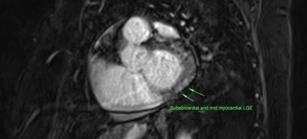
| 00:58, 1 August 2017 | Lymphocytic-myocarditis.jpg (file) |  |
77 KB | 1 | |
| 01:00, 1 August 2017 | 270780927951f0155ba941fe2264d1 big gallery.jpg (file) |  |
34 KB | 1 | |
| 12:32, 1 August 2017 | 386a52a8b0dc687cee574b7eaa00f9 jumbo.jpg (file) |  |
75 KB | 1 | |
| 12:35, 1 August 2017 | Medullary-bone-infarct-lupus-on-corticosteroids.JPG (file) |  |
55 KB | 1 | |
| 12:37, 1 August 2017 | Lupus-osteonecrosis-1.JPG (file) |  |
108 KB | 1 | |
| 12:47, 1 August 2017 | C4c3e9187bdede3c1e728e2eb0550a big gallery.jpeg (file) |  |
62 KB | Severe generalised fluid overload (pleural effusions, pericardial effusion, large volume ascites, subcutaneous oedema). Right renal vein thrombosis. | 1 |
| 13:04, 1 August 2017 | Osteoprosis mri.jpg (file) |  |
77 KB | 1 | |
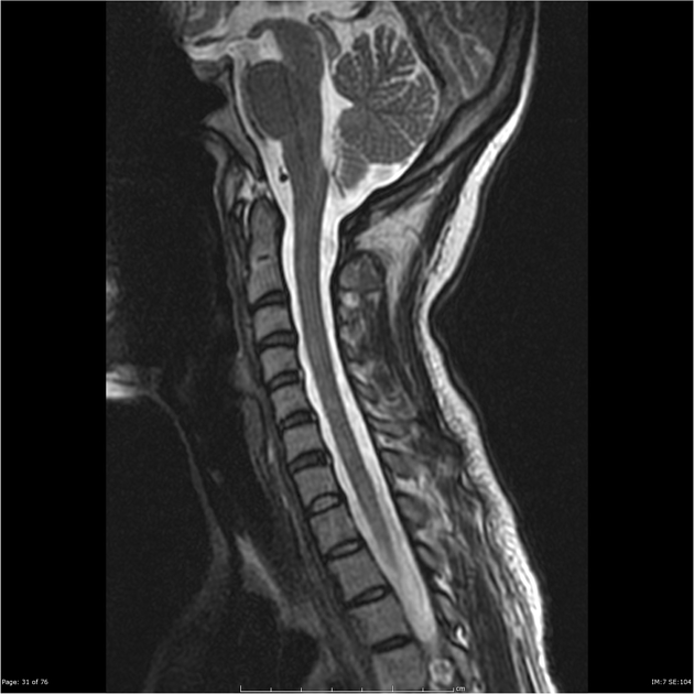
| 13:04, 1 August 2017 | Myelitis.jpg (file) |  |
125 KB | 1 | |
| 13:05, 1 August 2017 | E4b7a4cbffd49c1f7937c04e6b7c17 big gallery.jpeg (file) |  |
48 KB | 1 | |
| 13:07, 1 August 2017 | Osteoporosis-steroid-induced.jpg (file) |  |
81 KB | 1 | |
| 13:09, 1 August 2017 | Autosplenectomy.jpg (file) |  |
63 KB | 1 | |
| 13:17, 1 August 2017 | Colonic-pseudo-obstruction-ogilvies-syndrome.jpg (file) |  |
75 KB | 1 | |
| 13:18, 1 August 2017 | Ogilvie-syndrome-1.jpg (file) |  |
62 KB | 1 | |
| 13:20, 1 August 2017 | Acute-pancreatitis-with-walled-off-pancreatic-necrosis.jpg (file) |  |
99 KB | 1 | |
| 13:26, 1 August 2017 | Acute-infarction-1.jpg (file) |  |
63 KB | 1 | |
| 13:27, 1 August 2017 | Subacute-middle-cerebral-artery-infarct.jpg (file) |  |
162 KB | 1 | |
| 13:30, 1 August 2017 | 11b6d7d362d132027d14f5a6a12236 big gallery.jpg (file) |  |
48 KB | 1 | |
| 13:31, 1 August 2017 | Secondary-raynauds-phenomenon.jpg (file) |  |
75 KB | 1 | |
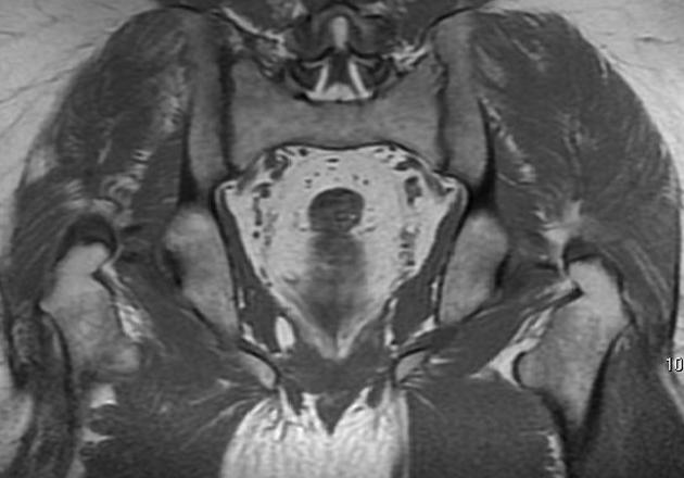
| 13:34, 1 August 2017 | 11f542413e3f946e7401ba6184820f big gallery.jpg (file) |  |
32 KB | STIR sequences showing hyperintense muscle signal involving the iliac muscles, bilateral, as well as gluteus maximus and medium, right side. | 1 |
| 13:49, 1 August 2017 | Extensor-carpi-ulnaris-tenosynovitis-1.jpg (file) |  |
37 KB | Global thickening with effusion is noted at sheath of extensor carpi ulnaris ( ECU ) tendon. Local hypervascularity also is present. There is normal fibrillar pattern at ECU tendon and no evidence of intratendinus hypoechoic areas /Tears. | 1 |
| 13:53, 1 August 2017 | A87cc2247174b9aad66f9bfe89bb1c big gallery.jpg (file) |  |
43 KB | Fluid is seen surrounding the tendon of Flexor hallucis longus tendon. It shows low signal on T1 and bright signal on T2 and well appreciated on fat suppressed images | 1 |
| 14:11, 1 August 2017 | Pericardial-effusion-sle.jpg (file) |  |
82 KB | 1 | |
| 14:13, 1 August 2017 | 2958c05a9bf1f2dc6b2f83ac4f8e81 jumbo.jpeg (file) |  |
66 KB | 1 | |
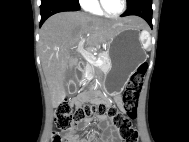
| 14:15, 1 August 2017 | 5e2515ac54c842fffa820c85e60acd big gallery.jpeg (file) |  |
34 KB | Ultrasound confirmed pericardial effusion with a mean thickness of approximately 30 mm, indicating a significant buildup of fluid in the pericardial sac (>20 mm: >500 ml) | 1 |
| 14:19, 1 August 2017 | 77a009fcd263e0e8def8755b5f47db big gallery.jpeg (file) |  |
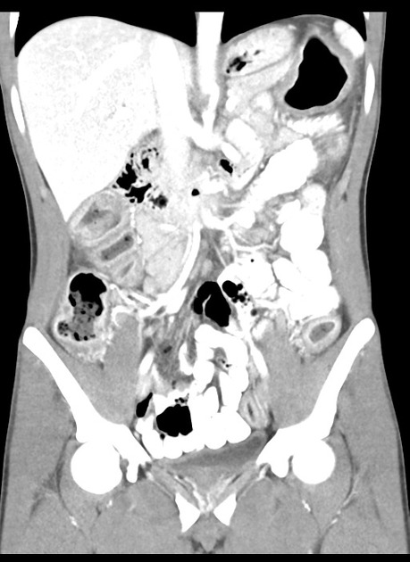
51 KB | Most of the small bowel is dilated with a transition point in the lower abdomen with swirling of the mesenteric vessels. The terminal 30 cm of small bowel is collapsed. There is poor enhancement of the wall of much of the small bowel, with gas in the s... | 1 |
| 14:20, 1 August 2017 | B95d2c43ad55d5df50c9b0323c3943 big gallery.jpg (file) |  |
86 KB | There is no pulmonary embolus identified. The pericardium is thickened with subtle stranding of the pericardial fat. Given the clinical setting, the appearance is most in keeping with pericarditis. | 1 |
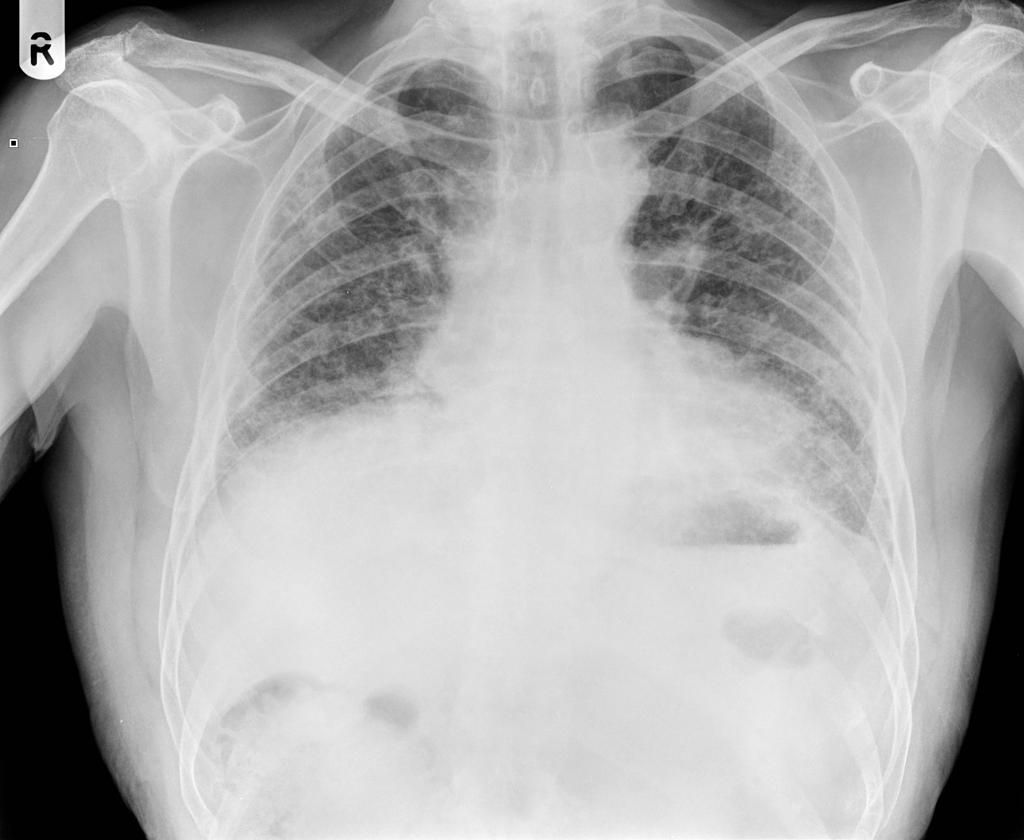
| 14:24, 1 August 2017 | Ddae1eb0ee1deec1a5d70aa127b608 jumbo.jpg (file) |  |
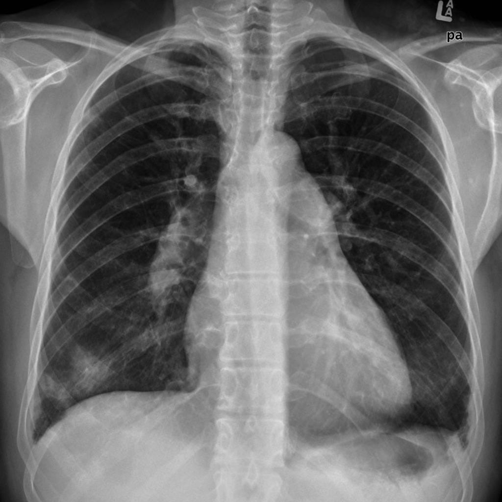
64 KB | Small volume lungs with bilateral increased interstitial markings, most pronounced peripherally, without a zonal predominence. | 1 |
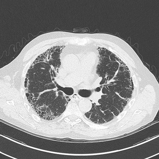
| 14:27, 1 August 2017 | E14c73a115e5aca00abc59216b76d2 big gallery.jpg (file) |  |
88 KB | Extensive honeycomb change throughout both lungs in a subpleural distribution with consequential tractional bronchiectasis. | 2 |
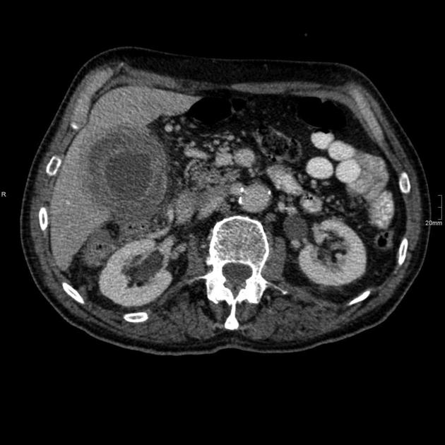
| 14:36, 1 August 2017 | Ca9623b810f6389be1e410a2827662 big gallery.jpeg (file) |  |
89 KB | The liver is mildly enlarged associated with periportal edema. Diffuse gallbladder wall thickening with no hyperdense luminal foci. Mild ascites is noted in Morison's pouch and in the pelvis. | 1 |
| 14:38, 1 August 2017 | 3fc11253ba09067fb09f32399ba387 big gallery.jpg (file) |  |
32 KB | Thickened and oedematous gall bladder wall with biliary sludge. | 1 |
| 14:40, 1 August 2017 | F22a56486ba4cfcd46a6577463ef02 big gallery.jpg (file) |  |
45 KB | Acute cholecystitis with intramural abscesses. | 1 |
| 14:41, 1 August 2017 | Acute-acalculous-cholecystitis-1.jpg (file) |  |
66 KB | acalculous-cholecystitis | 1 |
| 14:43, 1 August 2017 | Acute-cholecystitis-3.jpg (file) |  |
49 KB | pericholecystic fluid, gall bladder wall thickening | 1 |