Uploads by Mmir
Jump to navigation
Jump to search
This special page shows all uploaded files.
| Date | Name | Thumbnail | Size | Description | Versions |
|---|---|---|---|---|---|
| 16:22, 27 September 2018 | 123ttt.png (file) |  |
19 KB | 1 | |
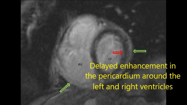
| 14:55, 1 August 2017 | 59f9b477529f76aad3615898fb5d6f big gallery.jpg (file) |  |
23 KB | In this cine grid sequence, the pericardium is not sliding past the myocardium as expected. This is shown by the lines extending through the pericardium and myocardium. They do not break at any point in the cardiac cycle. | 1 |
| 00:03, 23 August 2017 | Pac.jpg (file) | 27 KB | 1 | ||
| 17:45, 13 June 2017 | Molluscumcontagiosum1.jpg (file) |  |
27 KB | 1 | |
| 22:25, 1 October 2017 | Bernard courtois.jpg (file) |  |
29 KB | 1 | |
| 15:25, 1 August 2017 | C13ada80fec4008d775777547a65ef big gallery.jpg (file) |  |
31 KB | Gross dilatation of the pancreatic duct (and gland atrophy) with subtle filling defect in the duct at the head extending into the accessory duct draining the uncinate process - easily seen as calcification on CT but not that apparent on MRI. Calcifica... | 1 |
| 23:53, 22 August 2017 | Ggh.gif (file) |  |
31 KB | 1 | |
| 14:38, 1 August 2017 | 3fc11253ba09067fb09f32399ba387 big gallery.jpg (file) |  |
32 KB | Thickened and oedematous gall bladder wall with biliary sludge. | 1 |
| 13:34, 1 August 2017 | 11f542413e3f946e7401ba6184820f big gallery.jpg (file) |  |
32 KB | STIR sequences showing hyperintense muscle signal involving the iliac muscles, bilateral, as well as gluteus maximus and medium, right side. | 1 |
| 14:15, 1 August 2017 | 5e2515ac54c842fffa820c85e60acd big gallery.jpeg (file) |  |
34 KB | Ultrasound confirmed pericardial effusion with a mean thickness of approximately 30 mm, indicating a significant buildup of fluid in the pericardial sac (>20 mm: >500 ml) | 1 |
| 01:00, 1 August 2017 | 270780927951f0155ba941fe2264d1 big gallery.jpg (file) |  |
34 KB | 1 | |
| 20:09, 22 August 2017 | 3.jpg (file) |  |
36 KB | 2 | |
| 20:10, 22 August 2017 | Jjjkjgh.jpeg (file) |  |
36 KB | 1 | |
| 13:49, 1 August 2017 | Extensor-carpi-ulnaris-tenosynovitis-1.jpg (file) |  |
37 KB | Global thickening with effusion is noted at sheath of extensor carpi ulnaris ( ECU ) tendon. Local hypervascularity also is present. There is normal fibrillar pattern at ECU tendon and no evidence of intratendinus hypoechoic areas /Tears. | 1 |
| 15:31, 29 October 2018 | Ulnar fracture.jpg (file) |  |
37 KB | 1 | |
| 15:14, 29 October 2018 | Ulnar stick fx.jpeg (file) |  |
37 KB | 1 | |
| 15:20, 1 August 2017 | Ec9e2a6a4c1d377bdee5f0ccbbf180 big gallery.jpg (file) |  |
38 KB | Mild Ventriculomegaly. Restriction on diffusion in the periventricular region along bilateral lateral ventricles. Minimal ependymal enhancement on contrast administration. MRI features likely representing ventriculitis. | 1 |
| 00:00, 23 August 2017 | Ggddh.gif (file) |  |
38 KB | 1 | |
| 15:22, 1 August 2017 | D1797f6a1a111e62b0f9ba00fc93bb big gallery.jpg (file) |  |
38 KB | The left retrobulbar optic nerve demonstrated high T2 signal and enhancement. | 1 |
| 13:53, 1 August 2017 | A87cc2247174b9aad66f9bfe89bb1c big gallery.jpg (file) |  |
43 KB | Fluid is seen surrounding the tendon of Flexor hallucis longus tendon. It shows low signal on T1 and bright signal on T2 and well appreciated on fat suppressed images | 1 |
| 14:40, 1 August 2017 | F22a56486ba4cfcd46a6577463ef02 big gallery.jpg (file) |  |
45 KB | Acute cholecystitis with intramural abscesses. | 1 |
| 15:12, 1 August 2017 | 2739a8754252754d33b8ee2d413894 big gallery.jpg (file) |  |
46 KB | Curved MIP shows critical stenosis in the proximal LAD | 1 |
| 13:05, 1 August 2017 | E4b7a4cbffd49c1f7937c04e6b7c17 big gallery.jpeg (file) |  |
48 KB | 1 | |
| 13:30, 1 August 2017 | 11b6d7d362d132027d14f5a6a12236 big gallery.jpg (file) |  |
48 KB | 1 | |
| 14:43, 1 August 2017 | Acute-cholecystitis-3.jpg (file) |  |
49 KB | pericholecystic fluid, gall bladder wall thickening | 1 |
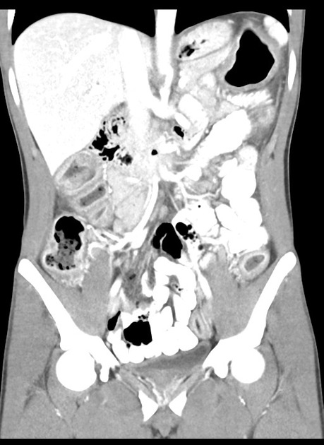
| 14:19, 1 August 2017 | 77a009fcd263e0e8def8755b5f47db big gallery.jpeg (file) |  |
51 KB | Most of the small bowel is dilated with a transition point in the lower abdomen with swirling of the mesenteric vessels. The terminal 30 cm of small bowel is collapsed. There is poor enhancement of the wall of much of the small bowel, with gas in the s... | 1 |
| 15:16, 1 August 2017 | 9763697dab2e8388d7a54b513a3e42 jumbo.jpg (file) |  |
52 KB | 1 | |
| 19:32, 14 August 2019 | NM.jpg (file) |  |
54 KB | 1 | |
| 12:35, 1 August 2017 | Medullary-bone-infarct-lupus-on-corticosteroids.JPG (file) |  |
55 KB | 1 | |
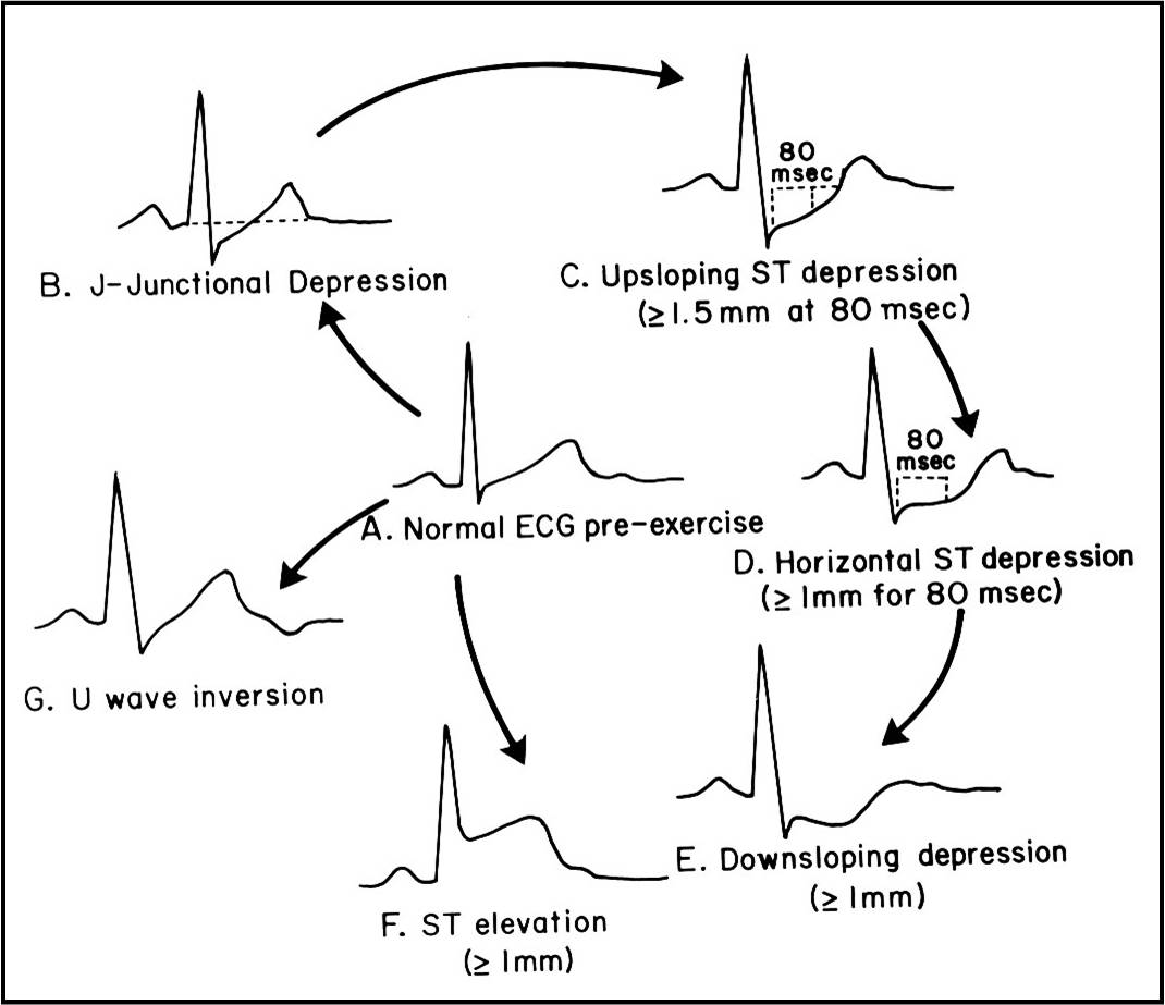
| 00:05, 23 August 2017 | Ecg 12lead006z.gif (file) |  |
56 KB | 1 | |
| 14:59, 1 August 2017 | 5069b7ce46c04b25f931f21561887b big gallery.jpg (file) |  |
56 KB | Multiple bilateral pulmonary emboli, largest focus in right lower lobe. Evidence of right heart strain with dilatation of right ventricle and atrium. Right basal consolidation is a possible site of infarction given distribution of emboli. Small right... | 1 |
| 19:34, 14 August 2019 | Nmmmm.jpg (file) |  |
56 KB | 1 | |
| 15:09, 1 August 2017 | Faa79e035d4c88b1029b3f6cd6e222 jumbo.jpeg (file) |  |
57 KB | Elevation of the right hemidiaphragm. | 1 |
| 15:16, 1 August 2017 | 9531e159d25c6beef968d6c1caf46c jumbo.jpg (file) |  |
59 KB | 1 | |
| 12:47, 1 August 2017 | C4c3e9187bdede3c1e728e2eb0550a big gallery.jpeg (file) |  |
62 KB | Severe generalised fluid overload (pleural effusions, pericardial effusion, large volume ascites, subcutaneous oedema). Right renal vein thrombosis. | 1 |
| 13:18, 1 August 2017 | Ogilvie-syndrome-1.jpg (file) |  |
62 KB | 1 | |
| 14:47, 1 August 2017 | Hepatomegaly-on-abdominal-x-ray.jpg (file) |  |
62 KB | Liver silhouette is enlarged, displacing bowel loops medially. | 1 |
| 13:26, 1 August 2017 | Acute-infarction-1.jpg (file) |  |
63 KB | 1 | |
| 13:09, 1 August 2017 | Autosplenectomy.jpg (file) |  |
63 KB | 1 | |
| 14:24, 1 August 2017 | Ddae1eb0ee1deec1a5d70aa127b608 jumbo.jpg (file) |  |
64 KB | Small volume lungs with bilateral increased interstitial markings, most pronounced peripherally, without a zonal predominence. | 1 |
| 00:39, 1 August 2017 | Thumbprinting ct.jpeg (file) |  |
65 KB | 1 | |
| 14:41, 1 August 2017 | Acute-acalculous-cholecystitis-1.jpg (file) |  |
66 KB | acalculous-cholecystitis | 1 |
| 14:13, 1 August 2017 | 2958c05a9bf1f2dc6b2f83ac4f8e81 jumbo.jpeg (file) |  |
66 KB | 1 | |
| 00:44, 1 August 2017 | Pleural effusion ct.jpg (file) |  |
67 KB | 1 | |
| 00:54, 1 August 2017 | Acute-hypersensitivity-pneumonitis.jpg (file) |  |
68 KB | 1 | |
| 23:39, 22 August 2017 | LA.gif (file) |  |
70 KB | 1 | |
| 23:50, 22 August 2017 | Jjj.gif (file) |  |
70 KB | 1 | |
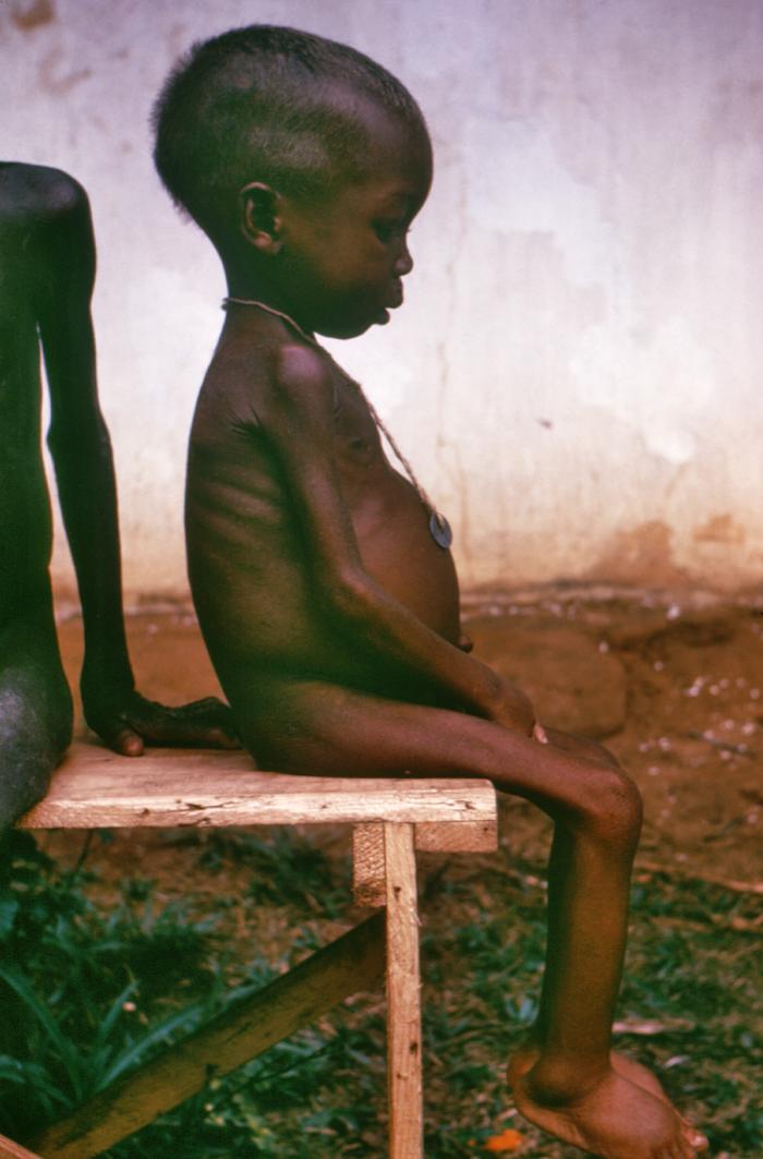
| 23:05, 19 September 2017 | Kwashiorkor.jpg (file) |  |
71 KB | 1 | |
| 00:15, 23 August 2017 | Jkhhk.jpg (file) |  |
72 KB | 1 | |
| 13:17, 1 August 2017 | Colonic-pseudo-obstruction-ogilvies-syndrome.jpg (file) |  |
75 KB | 1 |