Uploads by Esther Lee
Jump to navigation
Jump to search
This special page shows all uploaded files.
| Date | Name | Thumbnail | Size | Description | Versions |
|---|---|---|---|---|---|
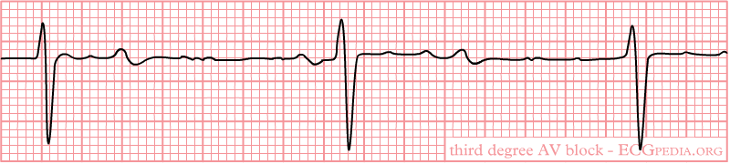
| 14:14, 16 October 2012 | 3rd Degree AV Block 1.png (file) | 11 KB | 1 | ||
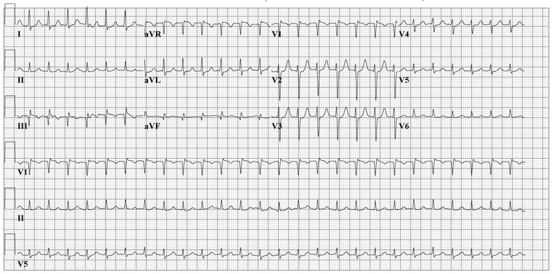
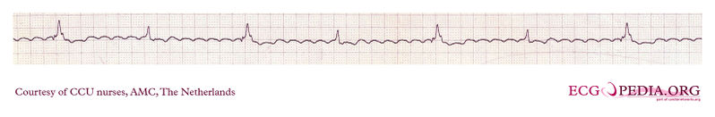
| 13:36, 16 October 2012 | AV nodal Reentrant Tachycardia 2.jpg (file) |  |
78 KB | 1 | |
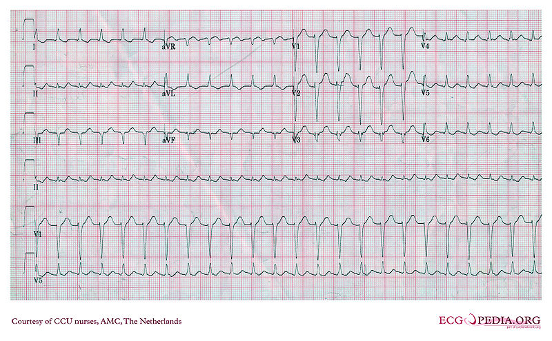
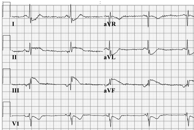
| 13:37, 16 October 2012 | AV nodal Reentrant Tachycardia 3.jpg (file) |  |
153 KB | 1 | |
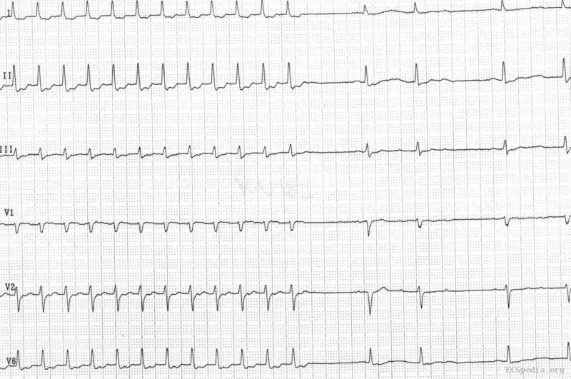
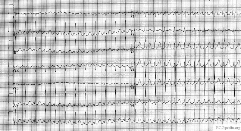
| 13:32, 16 October 2012 | AVnodalReentrantTachycardia1.jpg (file) |  |
130 KB | 1 | |
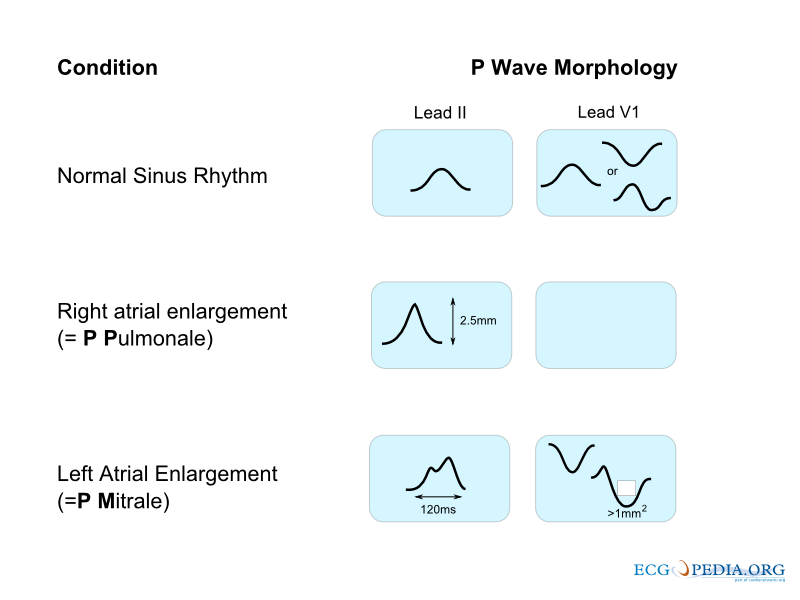
| 15:34, 15 October 2012 | AbnormalPWave.png (file) |  |
80 KB | 1 | |
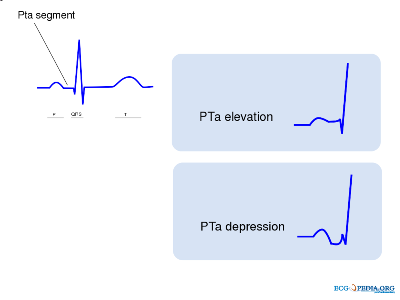
| 15:45, 15 October 2012 | AbnormalPWave2.png (file) |  |
38 KB | 1 | |
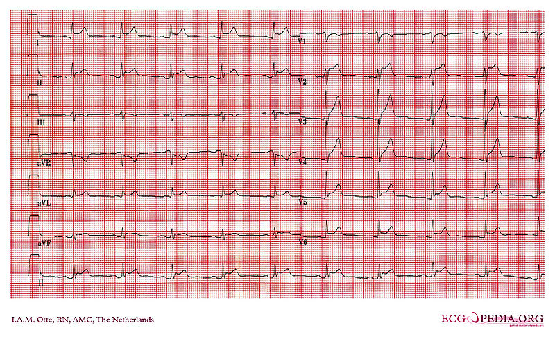
| 18:38, 15 October 2012 | AcutePericarditis.jpg (file) |  |
229 KB | 1 | |
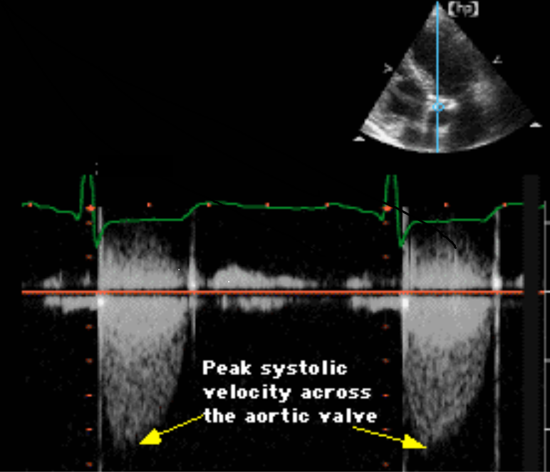
| 20:50, 2 October 2012 | Aortic stenosis echocardiography.png (file) |  |
208 KB | 1 | |
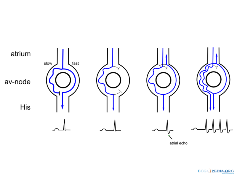
| 13:16, 16 October 2012 | ArrhythmiaPath.png (file) |  |
50 KB | 1 | |
| 16:00, 15 October 2012 | AtrialFibrillation2to1Conduction.jpg (file) |  |
108 KB | 1 | |
| 16:03, 15 October 2012 | AtrialFibrillationStrip.jpg (file) | 18 KB | 1 | ||
| 14:35, 16 October 2012 | Atrial Infarction 1.png (file) |  |
127 KB | 1 | |
| 16:40, 15 October 2012 | BrugadaSyndromeType2.jpg (file) |  |
41 KB | 1 | |
| 16:44, 15 October 2012 | BrugadaSyndromeType2 1.png (file) |  |
307 KB | 1 | |
| 17:00, 15 October 2012 | Brugada syndrome type1 example1.png (file) |  |
310 KB | 1 | |
| 17:01, 15 October 2012 | Brugada syndrome type1 example2.png (file) |  |
308 KB | 1 | |
| 17:02, 15 October 2012 | Brugada syndrome type1 example3.png (file) |  |
172 KB | 1 | |
| 17:17, 15 October 2012 | Brugada syndrome type1 example4.png (file) |  |
306 KB | 1 | |
| 17:18, 15 October 2012 | Brugada syndrome type1 example5.png (file) |  |
292 KB | 1 | |
| 20:37, 24 September 2012 | Complicated silicosis.jpg (file) |  |
46 KB | 1 | |
| 13:37, 15 October 2012 | CorrectedQT.png (file) | 754 bytes | 1 | ||
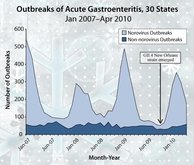
| 16:16, 20 November 2012 | Dsnorovirus 379px.jpg (file) |  |
21 KB | 1 | |
| 16:14, 20 November 2012 | Dsnorovirus 626px.jpg (file) |  |
55 KB | 1 | |
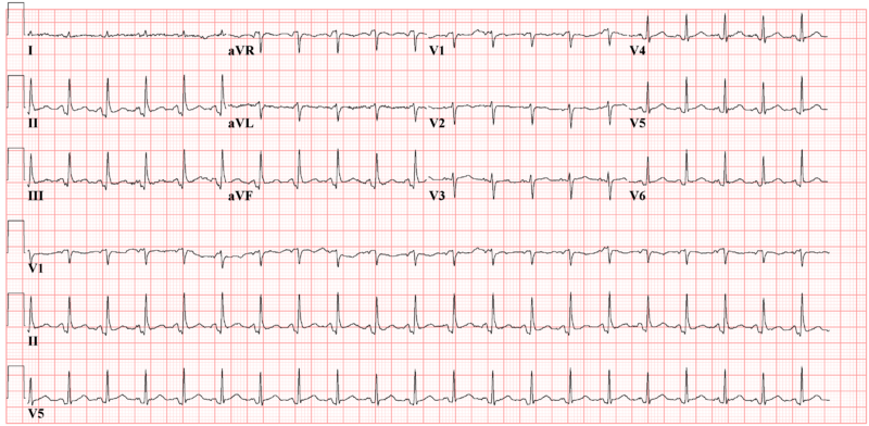
| 14:41, 16 October 2012 | Ectopic Atrial Rhythm 1.png (file) |  |
127 KB | Ectopic atrial rhythm due to atrial infarction | 1 |
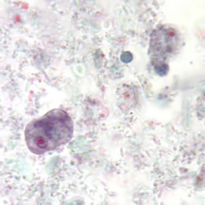
| 17:18, 19 November 2012 | Enana troph trich2.jpg (file) |  |
16 KB | 1 | |
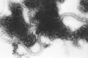
| 20:52, 26 September 2012 | Human RSV.jpg (file) |  |
16 KB | 1 | |
| 16:39, 19 November 2012 | Intestinal amebae.gif (file) |  |
72 KB | 1 | |
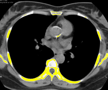
| 20:08, 2 October 2012 | Plain ct.jpg (file) |  |
28 KB | Plain CT showing an impressive almost circular supravalvular aortic calcification of the aortic wall with extension of calcifications into the left main stem. The area of calcification is shown in yellow. | 1 |
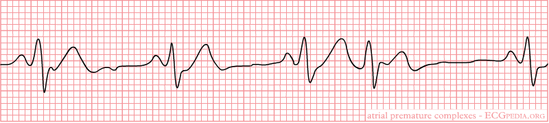
| 16:45, 16 October 2012 | Premature Atrial Contraction 6.png (file) | 9 KB | 1 | ||
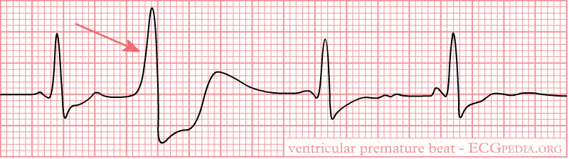
| 16:23, 16 October 2012 | Premature Ventricular 1.png (file) |  |
10 KB | 1 | |
| 16:35, 16 October 2012 | Premature Ventricular 2.png (file) | 9 KB | 1 | ||
| 18:48, 15 October 2012 | PtaDepressionPericarditis.png (file) |  |
126 KB | 1 | |
| 16:22, 15 October 2012 | RightVentricularMyocardialInfarction.jpg (file) |  |
78 KB | 1 | |
| 20:36, 24 September 2012 | SilicosisXRay.jpg (file) |  |
43 KB | 1 | |
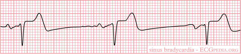
| 13:55, 15 October 2012 | Sinusbradycardia.png (file) |  |
221 KB | 2 | |
| 14:05, 15 October 2012 | Sinusbradycardia2.png (file) | 9 KB | 1 | ||
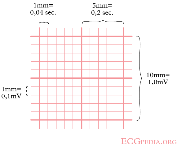
| 15:12, 15 October 2012 | Square counting method.png (file) |  |
6 KB | 2 | |
| 15:20, 15 October 2012 | TheMarkerMethod.png (file) |  |
13 KB | 1 | |
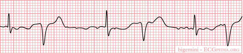
| 16:15, 16 October 2012 | Ventricular Bigemini 1.png (file) | 9 KB | 1 | ||
| 15:48, 16 October 2012 | Ventricular Tachycardia 1.jpg (file) |  |
75 KB | 2 | |
| 15:44, 16 October 2012 | Ventricular Tachycardia 2.png (file) |  |
37 KB | 1 | |
| 15:50, 16 October 2012 | Ventricular Tachycardia 3.jpg (file) |  |
197 KB | 1 | |
| 15:53, 16 October 2012 | Ventricular Tachycardia 4.jpg (file) |  |
205 KB | 1 | |
| 14:06, 16 October 2012 | Ventriculophasic Reflex.jpg (file) | 53 KB | 1 | ||
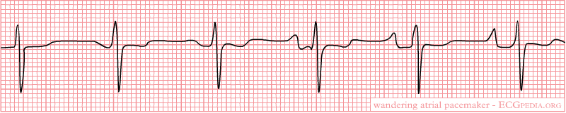
| 14:18, 16 October 2012 | Wandering Atrial Pacemaker 1.png (file) | 9 KB | 1 |