Uploads by Mmir
Jump to navigation
Jump to search
This special page shows all uploaded files.
| Date | Name | Thumbnail | Size | Description | Versions |
|---|---|---|---|---|---|
| 13:31, 1 August 2017 | Secondary-raynauds-phenomenon.jpg (file) |  |
75 KB | 1 | |
| 12:32, 1 August 2017 | 386a52a8b0dc687cee574b7eaa00f9 jumbo.jpg (file) |  |
75 KB | 1 | |
| 00:58, 1 August 2017 | Lymphocytic-myocarditis.jpg (file) |  |
77 KB | 1 | |
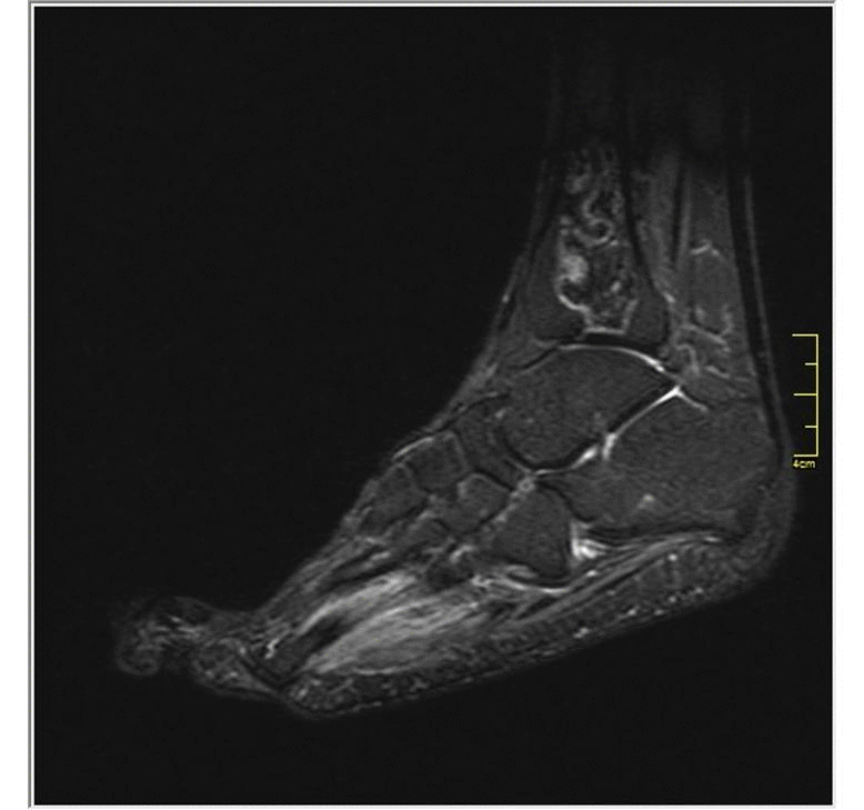
| 13:04, 1 August 2017 | Osteoprosis mri.jpg (file) |  |
77 KB | 1 | |
| 13:07, 1 August 2017 | Osteoporosis-steroid-induced.jpg (file) |  |
81 KB | 1 | |
| 14:11, 1 August 2017 | Pericardial-effusion-sle.jpg (file) |  |
82 KB | 1 | |
| 00:45, 1 August 2017 | Pleural effusion graphy.jpg (file) |  |
83 KB | 1 | |
| 16:33, 6 May 2018 | 3392802 orig.jpg (file) |  |
85 KB | 1 | |
| 16:03, 1 May 2018 | Upper anatomy.jpeg (file) |  |
85 KB | 1 | |
| 14:20, 1 August 2017 | B95d2c43ad55d5df50c9b0323c3943 big gallery.jpg (file) |  |
86 KB | There is no pulmonary embolus identified. The pericardium is thickened with subtle stranding of the pericardial fat. Given the clinical setting, the appearance is most in keeping with pericarditis. | 1 |
| 14:27, 1 August 2017 | E14c73a115e5aca00abc59216b76d2 big gallery.jpg (file) |  |
88 KB | Extensive honeycomb change throughout both lungs in a subpleural distribution with consequential tractional bronchiectasis. | 2 |
| 14:36, 1 August 2017 | Ca9623b810f6389be1e410a2827662 big gallery.jpeg (file) |  |
89 KB | The liver is mildly enlarged associated with periportal edema. Diffuse gallbladder wall thickening with no hyperdense luminal foci. Mild ascites is noted in Morison's pouch and in the pelvis. | 1 |
| 15:02, 1 August 2017 | A015828ba66db9da16b5a57d0cb2c6 jumbo.jpeg (file) |  |
92 KB | steroid-induced osteoprosis | 1 |
| 00:56, 1 August 2017 | Hypersensitivity-pneumonitis.jpg (file) |  |
94 KB | 1 | |
| 00:51, 1 August 2017 | Pulmonary hypertension graphy.jpg (file) |  |
97 KB | 1 | |
| 13:20, 1 August 2017 | Acute-pancreatitis-with-walled-off-pancreatic-necrosis.jpg (file) |  |
99 KB | 1 | |
| 17:04, 25 September 2017 | Mohsin gif.gif (file) |  |
101 KB | 1 | |
| 23:47, 22 August 2017 | 84 Acute inf STEMI.jpg (file) |  |
105 KB | 1 | |
| 14:58, 1 August 2017 | 8272710ac07076fc011a7e0ad2fc7a jumbo.jpg (file) |  |
108 KB | Patchy opacification at the right base. No pneumothorax. Normal heart size. No bony abnormalities. | 1 |
| 12:37, 1 August 2017 | Lupus-osteonecrosis-1.JPG (file) |  |
108 KB | 1 | |
| 00:50, 1 August 2017 | Pulmonary-arterial-hypertension-7.jpg (file) |  |
115 KB | 1 | |
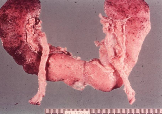
| 23:15, 21 August 2017 | Liver hydatic gross.jpg (file) |  |
120 KB | 1 | |
| 13:04, 1 August 2017 | Myelitis.jpg (file) |  |
125 KB | 1 | |
| 00:38, 1 August 2017 | Thumbprinting xray.jpeg (file) |  |
132 KB | 1 | |
| 23:36, 13 June 2017 | Molluscaklein.jpg (file) |  |
142 KB | 2 | |
| 23:10, 28 July 2017 | Lupus final.jpg (file) |  |
154 KB | 1 | |
| 00:33, 1 August 2017 | Lupus2.jpg (file) |  |
154 KB | 1 | |
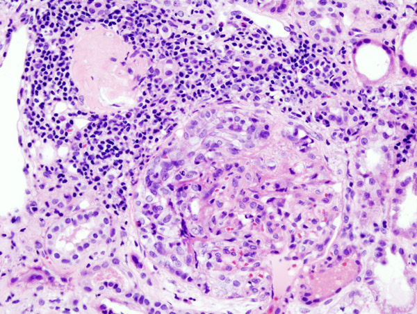
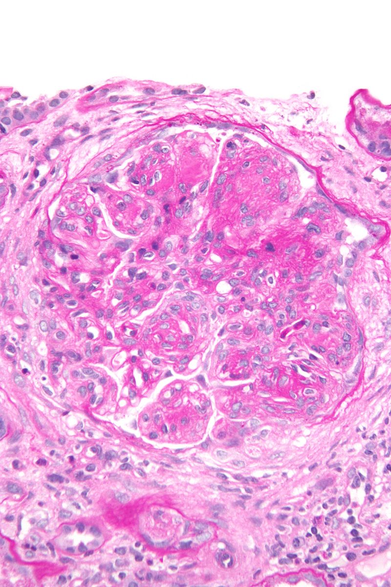
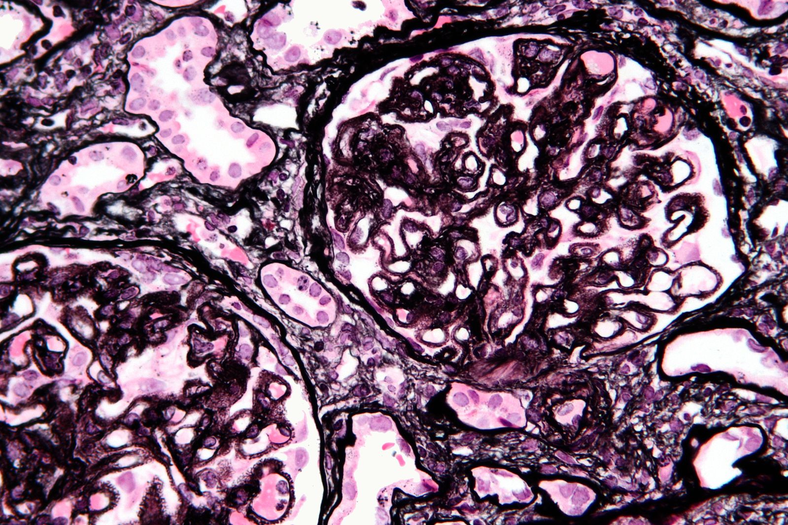
| 22:55, 31 July 2017 | Crescentic glomerulonephritis (2).jpg (file) | .jpg) |
160 KB | 1 | |
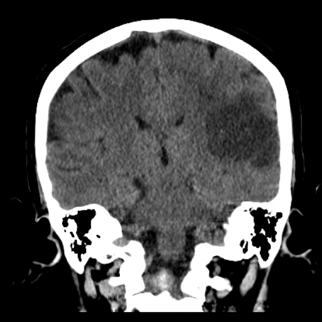
| 13:27, 1 August 2017 | Subacute-middle-cerebral-artery-infarct.jpg (file) |  |
162 KB | 1 | |
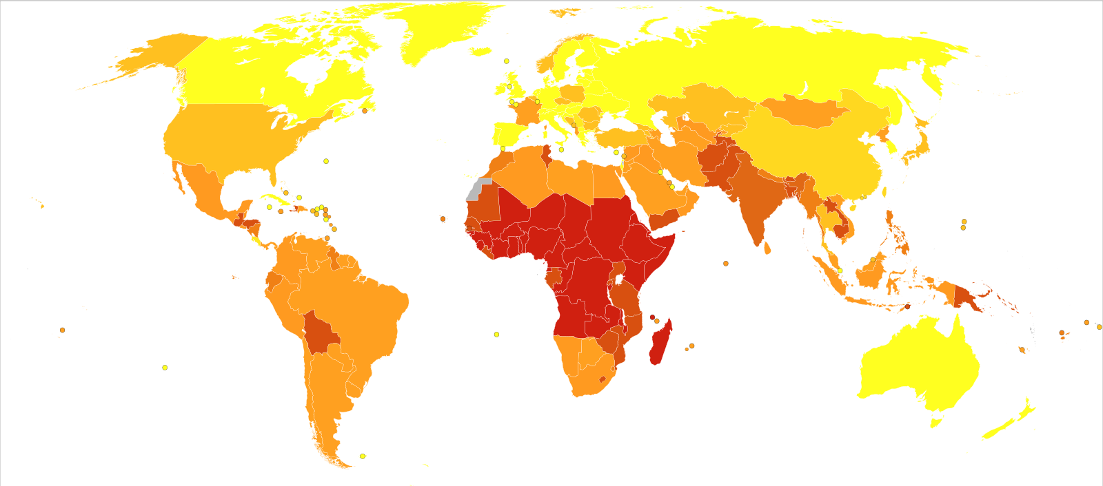
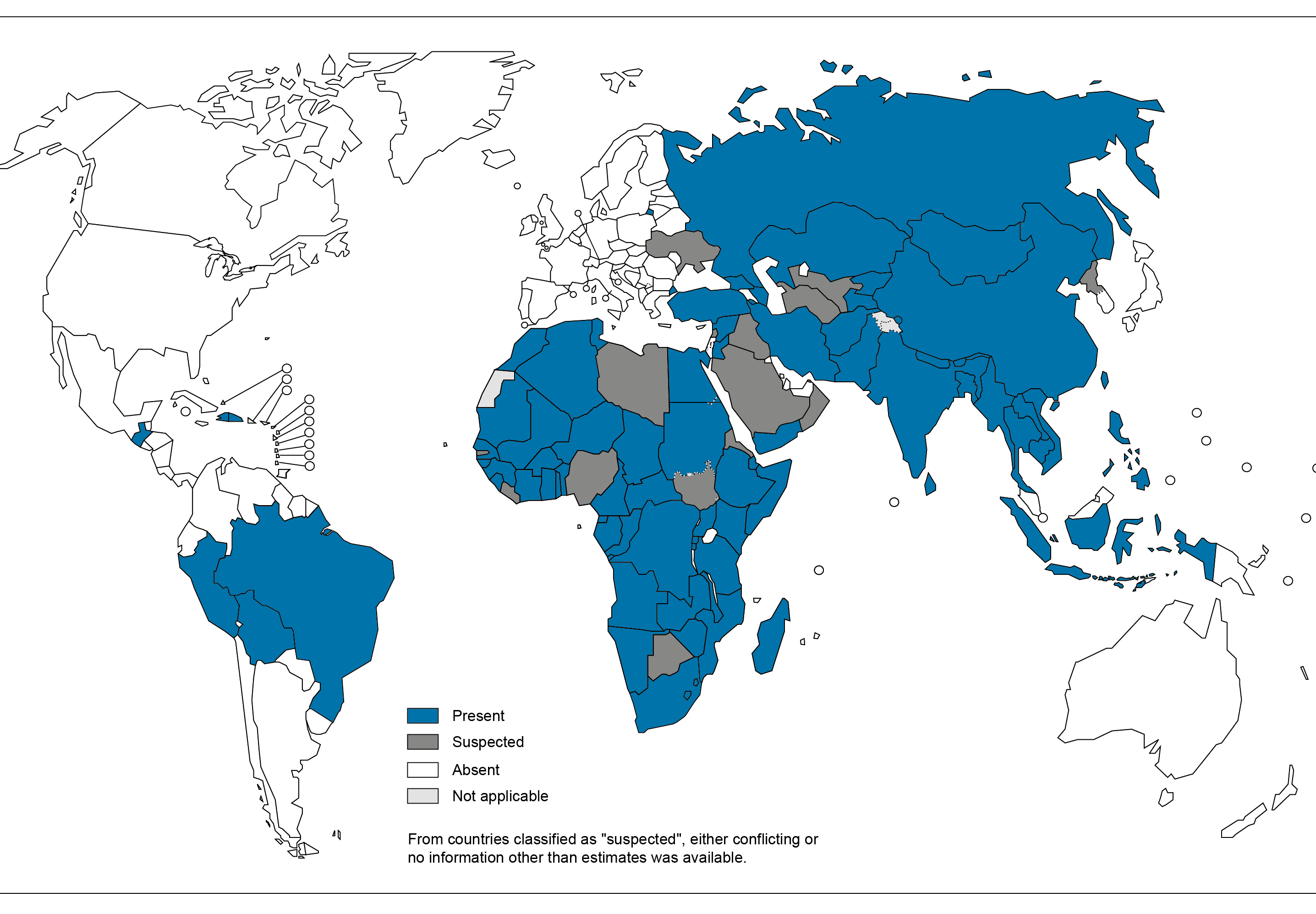
| 22:49, 19 September 2017 | Protein-energy malnutrition world map-DALYs per million persons-WHO2012.svg.png (file) |  |
192 KB | 1 | |
| 22:59, 31 July 2017 | Membranoproliferative glomerulonephritis - very high mag.jpg (file) |  |
192 KB | 1 | |
| 12:23, 13 September 2017 | Webp.g-gifmaker (2).gif (file) | .gif) |
197 KB | 1 | |
| 16:58, 7 March 2018 | .r.png (file) |  |
205 KB | 1 | |
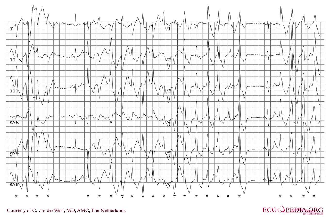
| 20:44, 9 January 2020 | Polymorphic vt.jpg (file) |  |
229 KB | 1 | |
| 23:55, 22 August 2017 | 80 RVH and LAE in mitral stenosis.jpg (file) |  |
282 KB | 1 | |
| 00:02, 23 August 2017 | 80 RVH and LAcxE in mitral stenosis.jpg (file) |  |
340 KB | 1 | |
| 21:55, 31 July 2017 | Vacuolar interface dermatitis - high mag.jpg (file) |  |
388 KB | 1 | |
| 22:56, 31 July 2017 | 1599px-Focal segmental glomerulosclerosis - high mag.jpg (file) |  |
389 KB | 1 | |
| 22:27, 1 October 2017 | Leonardo.jpg (file) |  |
389 KB | 1 | |
| 15:04, 1 August 2017 | 56dce39ec7860a50a4bf060db455b7 jumbo.jpeg (file) |  |
402 KB | Right upper lobe ground glass alveolar opacity and air bronchogram sign due to mitral regurgitation and secondary pulmonary edema | 1 |
| 22:57, 31 July 2017 | Membranous nephropathy - mpas - very high mag.jpg (file) |  |
406 KB | 1 | |
| 23:32, 20 August 2017 | Echinococcus granulosus scolex.jpg (file) |  |
406 KB | 1 | |
| 23:44, 21 August 2017 | Webp.net-gifmaker (37).gif (file) | .gif) |
409 KB | 1 | |
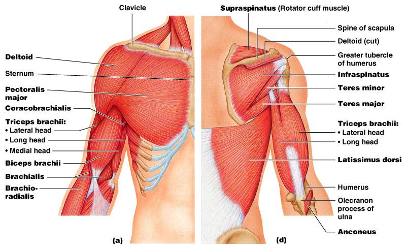
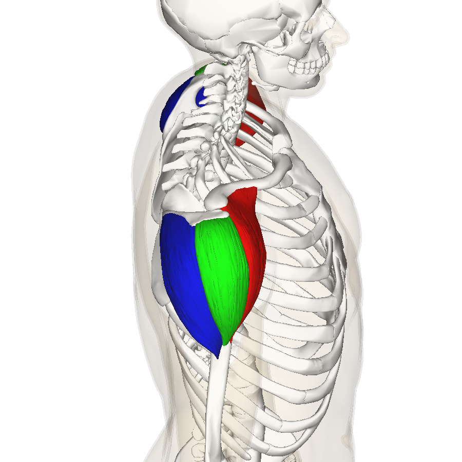
| 16:17, 1 May 2018 | Deltoid muscle top10.png (file) |  |
414 KB | 1 | |
| 00:37, 21 August 2017 | Webp.net-gifmaker (32).gif (file) | .gif) |
417 KB | 1 | |
| 22:38, 22 August 2017 | Webp.net-gifmjyfssaker (7).gif (file) | .gif) |
463 KB | 1 | |
| 20:12, 23 January 2019 | Renalslide 32.png (file) |  |
481 KB | 1 | |
| 18:22, 27 September 2017 | Rabies11.png (file) |  |
497 KB | 2 | |
| 23:35, 21 August 2017 | Webp.net-gifmaker (36).gif (file) | .gif) |
503 KB | 1 | |
| 14:12, 9 August 2017 | Webp.net-gifmaker (28).gif (file) | .gif) |
544 KB | 1 |