Polycystic ovary syndrome ultrasound
|
Polycystic ovary syndrome Microchapters |
|
Differentiating Polycystic ovary syndrome from other Diseases |
|---|
|
Diagnosis |
|
Treatment |
|
Case Studies |
|
Polycystic ovary syndrome ultrasound On the Web |
|
American Roentgen Ray Society Images of Polycystic ovary syndrome ultrasound |
|
Risk calculators and risk factors for Polycystic ovary syndrome ultrasound |
Editor-In-Chief: C. Michael Gibson, M.S., M.D. [1]; Associate Editor(s)-in-Chief:
Please help WikiDoc by adding content here. It's easy! Click here to learn about editing.
Overview
The Rotterdam 2003 criteria include the use of ultrasound as a diagnostic tool. The use of ultrasonography in the diagnosis of PCOS must be tempered by an awareness of the broad spectrum of women with ultrasonographic findings characteristic of polycystic ovaries. The typical polycystic-appearing ovary may emerge in a nonspecific fashion when a state of anovulation of any cause persists for any length of time (see Fig. 17-29 ). 209 Thus, the polycystic-appearing ovary is the result of a functional derangement but not a specific central or local defect.
Ultrasound
Ultrasonography
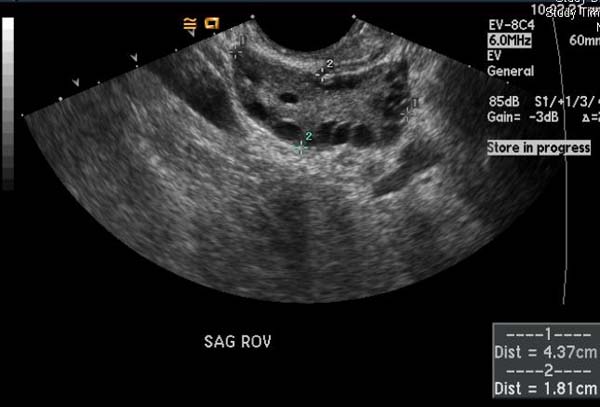
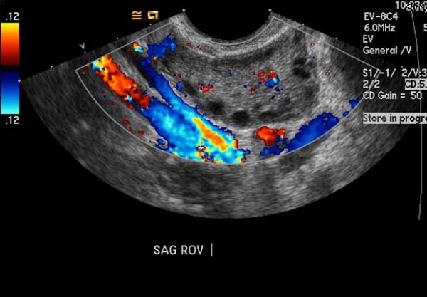
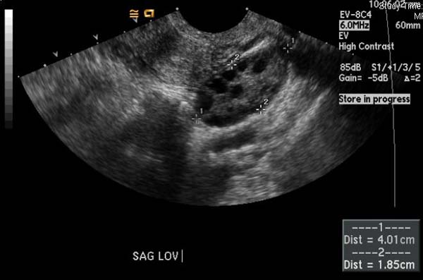
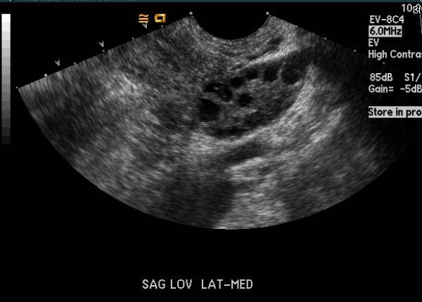
Typical ultrasonography findings in patients with PCOS are as follows:[1]
- Two- to 5-fold ovarian enlargement; ovarian volume >10 cm3
- Thickened stroma (tunica albuginea)
- Thecal hyperplasia with an increase in stromal content
- Multiple (12+) subcapsular follicles ranging from 2 to 9 mm in diameter in a state of arrested development ('pearl necklace' appearance); most are atretic and not necessarily cystic
- A discrete androgen-producing tumor in the ovary may be present in 5% or fewer patients
- The endometrium may be hyperplastic despite low estrogen production by the follicles; this is probably due to high estrone production from the increased circulating androgens and lack of opposition by progesterone
- Clomiphene may alter results due to ovarian stimulation, resulting in multiple ovarian cysts
-
Polycystic ovary syndrome
-
Polycystic ovary syndrome
-
Polycystic ovary syndrome
-
Polycystic ovary syndrome


References
- ↑ Kenigsberg LE, Agarwal C, Sin S, Shifteh K, Isasi CR, Crespi R, Ivanova J, Coupey SM, Heptulla RA, Arens R (2015). "Clinical utility of magnetic resonance imaging and ultrasonography for diagnosis of polycystic ovary syndrome in adolescent girls". Fertil. Steril. 104 (5): 1302–9.e1–4. doi:10.1016/j.fertnstert.2015.08.002. PMC 4630153. PMID 26354095.