Pituitary adenoma CT: Difference between revisions
No edit summary |
|||
| Line 7: | Line 7: | ||
==Key CT Scan Findings in Pituitary Adenoma== | ==Key CT Scan Findings in Pituitary Adenoma== | ||
===Macroadenoma=== | ===Macroadenoma=== | ||
*A | *A large [[suprasellar]] mass that typically characterized by:<ref name=radio>Pituitary Macroadenoma. Dr Bruno Di Muzio and Dr Yuranga Weerakkody. Radiopaedia.org 2015. http://radiopaedia.org/articles/pituitary-macroadenoma-1</ref> | ||
:*An [[attenuation]] similar to that of the brain (30-40 HU) | |||
:*Moderate [[contrast]] enhancement | |||
:*Invasion of the surrounding structures | |||
*Calcification and hemorrhage are rarely seen.<ref name=Radio>age courtesy of Dr Gagandeep Choudhary. http://www.radiopaedia.org Radiopaedia (http://radiopaedia.org/cases/pituitary-macroadenoma). http://radiopaedia.org/licence Creative Commons BY-SA-NC.</ref> | |||
<gallery> | <gallery> | ||
Image: | Image: | ||
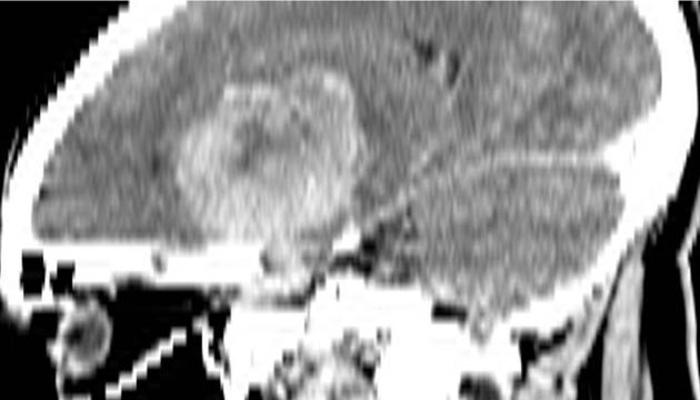
Pituitary adenoma.jpg|Image courtesy of Dr Gagandeep Choudhary. http://www.radiopaedia.org Radiopaedia | Pituitary adenoma.jpg|Large hyperdense sellar mass extending into the suprasellar region and causing displacement of surrounding structures. Image courtesy of Dr Gagandeep Choudhary. http://www.radiopaedia.org Radiopaedia (http://radiopaedia.org/cases/pituitary-macroadenoma). http://radiopaedia.org/licence Creative Commons BY-SA-NC. | ||
Pituitary adenoma.1.jpg|Image courtesy of Dr Gagandeep Choudhary. http://www.radiopaedia.org Radiopaedia | Pituitary adenoma.1.jpg|Large hyperdense sellar mass extending into the suprasellar region and causing displacement of surrounding structures. Image courtesy of Dr Gagandeep Choudhary. http://www.radiopaedia.org Radiopaedia (http://radiopaedia.org/cases/pituitary-macroadenoma). http://radiopaedia.org/licence Creative Commons BY-SA-NC. | ||
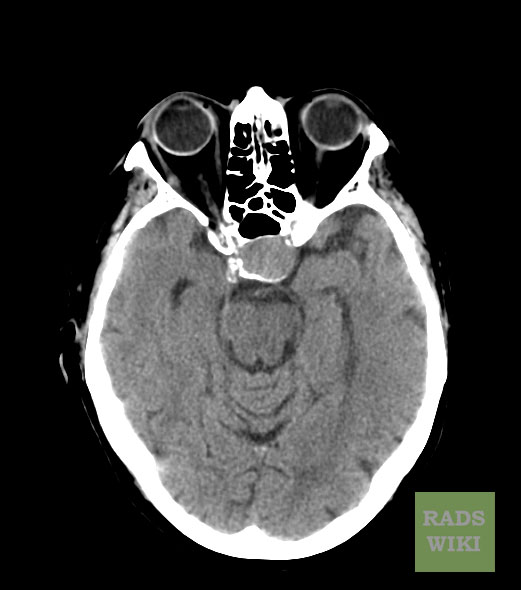
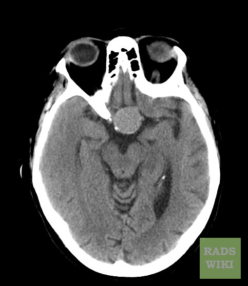
Pituitary-macroadenoma-CT-01.jpg| | Pituitary-macroadenoma-CT-01.jpg|There is a well defined round isodense lesion noted in the pituitary fossa, the lesion is widening the sella.http://www.radiopaedia.org Radiopaedia (http://radiopaedia.org/cases/pituitary-adenoma). | ||
Image:Pituitary-macroadenoma-CT-02.jpg|There is a well defined round isodense lesion noted in the pituitary fossa, the lesion is widening the sella.http://www.radiopaedia.org Radiopaedia (http://radiopaedia.org/cases/pituitary-adenoma). | |||
Image:Pituitary-macroadenoma-CT-02.jpg | |||
</gallery> | </gallery> | ||
Revision as of 20:42, 29 September 2015
|
Pituitary adenoma Microchapters |
|
Diagnosis |
|---|
|
Treatment |
|
Case Studies |
|
Pituitary adenoma CT On the Web |
|
American Roentgen Ray Society Images of Pituitary adenoma CT |
Editor-In-Chief: C. Michael Gibson, M.S., M.D. [1] Associate Editor(s)-in-Chief: Ahmad Al Maradni, M.D. [2]
Overview
Key CT Scan Findings in Pituitary Adenoma
Macroadenoma
- A large suprasellar mass that typically characterized by:[1]
- An attenuation similar to that of the brain (30-40 HU)
- Moderate contrast enhancement
- Invasion of the surrounding structures
- Calcification and hemorrhage are rarely seen.[2]
-
Large hyperdense sellar mass extending into the suprasellar region and causing displacement of surrounding structures. Image courtesy of Dr Gagandeep Choudhary. http://www.radiopaedia.org Radiopaedia (http://radiopaedia.org/cases/pituitary-macroadenoma). http://radiopaedia.org/licence Creative Commons BY-SA-NC.
-
There is a well defined round isodense lesion noted in the pituitary fossa, the lesion is widening the sella.http://www.radiopaedia.org Radiopaedia (http://radiopaedia.org/cases/pituitary-adenoma).
-
There is a well defined round isodense lesion noted in the pituitary fossa, the lesion is widening the sella.http://www.radiopaedia.org Radiopaedia (http://radiopaedia.org/cases/pituitary-adenoma).


Microadenoma
- Historically, before the advent of MRI, the pituitary was imaged with CT scan.
- Although CT scan is able to detect up to 80-90% of microadenomas (5-10mm in size), it has less sensitivity to smaller adenomas (less than 5mm in size).[1]
References
- ↑ 1.0 1.1 Pituitary Macroadenoma. Dr Bruno Di Muzio and Dr Yuranga Weerakkody. Radiopaedia.org 2015. http://radiopaedia.org/articles/pituitary-macroadenoma-1
- ↑ age courtesy of Dr Gagandeep Choudhary. http://www.radiopaedia.org Radiopaedia (http://radiopaedia.org/cases/pituitary-macroadenoma). http://radiopaedia.org/licence Creative Commons BY-SA-NC.