|
|
| Line 14: |
Line 14: |
| }} | | }} |
| {{Pericardial effusion}} | | {{Pericardial effusion}} |
| {{CMG}}; '''Associate Editor-In-Chief:''' {{CZ}} | | {{CMG}}; '''Associate Editor-In-Chief:''' {{CZ}}; [[Varun Kumar]], M.B.B.S |
| | |
| '''See also: [[pericarditis]], [[constrictive pericarditis]] and [[cardiac tamponade]]'''
| |
| | |
| ==Overview==
| |
| == Differential Diagnosis of {{PAGENAME}}==
| |
| | |
| === Serous ===
| |
| | |
| * [[Acute pancreatitis]]
| |
| * [[Chemotherapeutics]]
| |
| * [[Chronic disease]]
| |
| * [[Cirrhosis]]
| |
| * [[Congestive heart failure]]
| |
| * [[Dressler's syndrome]]
| |
| * [[Hypoalbuminemia]]
| |
| * [[Hypothyroidism]]
| |
| * [[Infection]]
| |
| * [[Irradiation]]
| |
| * [[Malnutrition]]
| |
| * [[Nephrotic Syndrome]]
| |
| | |
| === Blood ===
| |
| | |
| * [[STEMI|Acute myocardial infarction]]
| |
| * [[Anticoagulants]]
| |
| * [[chest trauma|Aortic rupture]]
| |
| * [[Cardiac catheterization]]
| |
| * [[Chemotherapeutics]]
| |
| * [[vitamin K|Coagulotherapy]]
| |
| * [[Heart surgery]]
| |
| * [[Neoplasm]]
| |
| * [[Perforation]]
| |
| * [[Trauma]]
| |
| * [[Uremia]]
| |
| | |
| === Lymph or chylous ===
| |
| | |
| * Benign obstruction of [[thoracic duct]]
| |
| * [[Idiopathic]]
| |
| * [[Neoplasm]]
| |
| | |
| === Metastatic tumor ===
| |
| | |
| * [[Breast cancer]]
| |
| * [[Leukemia]]
| |
| * [[Lung cancer]]
| |
| * [[Lymphoma]]
| |
| * [[Pericarditis]]
| |
| | |
| === Miscellaneous ===
| |
| | |
| * [[Cardiomyopathy]]
| |
| * [[Systemic Lupus Erythematosus]]
| |
| | |
| === Infectious===
| |
| | |
| * [[Viral]]
| |
| * [[Pyogenic]]
| |
| * [[Tuberculosis | Tuberculous]]
| |
| * [[Fungal]]
| |
| * [[infection|Other infections]] ([[syphilitic]], [[protozoal]], [[parasitic]])
| |
| * [[Pericarditis]]
| |
| | |
| ===Noninfectious===
| |
| | |
| * [[Idiopathic]]
| |
| * [[Uremia]]: [[Kidney failure]] with excessive blood levels of urea nitrogen
| |
| * [[Heart surgery]]<ref>[http://www.mayoclinic.com/health/pericardial-effusion/HQ01198 Pericardial effusion:What are the symptoms?], Dr. Martha Grogan M.D.</ref>
| |
| * [[Neoplasia]] that has spread to the [[pericardium]]
| |
| * [[Acute myocardial infarction]]: [[Dressler's syndrome|Post myocardial infarction pericarditis]] ([[Dressler's syndrome]])
| |
| * [[Radiation therapy|Postirradiation]]
| |
| * [[Aortic dissection]] (with leakage into [[pericardial sac]])
| |
| * [[Trauma]]
| |
| * [[Sarcoidosis]]
| |
| * [[Pericarditis]]
| |
| | |
| ===Hypersensitivity or autoimmunity related===
| |
| | |
| * [[Rheumatic fever]]
| |
| * [[Collagen vascular disease]]
| |
| * [[Drug-induced]]
| |
| * [[Inflammatory disease]], such as [[lupus]]
| |
| * [[Pericarditis]]
| |
| | |
| ==Types==
| |
| | |
| ===Transudative===
| |
| | |
| * [[congestive heart failure]]
| |
| * [[myxoedema]]
| |
| * [[nephrotic syndrome]]
| |
| | |
| ===Exudative===
| |
| | |
| * [[tuberculosis]],
| |
| * spread from [[empyema]]
| |
| | |
| ===Hemorrhagic===
| |
| | |
| * [[chest trauma]]
| |
| * [[aneurysm|rupture of aneurym]]s
| |
| * [[neoplasm|malignant effusion]]
| |
| | |
| ==Symptoms==
| |
| | |
| [[Chest pain]], pressure symptoms. A small effusion may have no symptoms.
| |
| | |
| Pericardial effusion is also present after a specific type of heart defect repair. An [[Atrial Septal Defect]] Secundum, or [[ASD]], when repaired will most likely produce a pericardial effusion due to one of the methods of repair. One repair method of an [[ASD]] is to take a piece of the peridcardial tissue and use it as a patch for the hole in the atrial cavity.
| |
|
| |
|
| | ==[[Pericardial effusion overview|Overview]]== |
| | ==[[Pericardial effusion differential diagnosis|Differential diagnosis]]== |
| | ==[[Pericardial effusion types|Types of pericardial effusion]]== |
| ==Diagnosis== | | ==Diagnosis== |
| | [[Pericardial effusion history and symptoms|Symptoms]] | [[Pericardial effusion physical examination|Physical examination]] | [[Pericardial effusion electrocardiogram|Electrocardiogram]] | [[Pericardial effusion chest x ray|Chest X-Ray]] | [[Pericardial effusion echocardiography|Echocardiography]] | [[Pericardial effusion CT and MRI|CT and MRI]] | [[Pericardial effusion cardiac catheterization|Cardiac catheterization]] |
|
| |
|
| ===EKG=== | | ==[[Pericardial effusion treatment overview|Treatment]]== |
| | |
| <gallery>
| |
| Image:Alternans.jpg|Pericardial Effusion
| |
| Image:Electrical Alternans.jpg|An ECG showing electrical alternans in a person with pericardial effusion.
| |
| </gallery>
| |
| | |
| <br clear="left"/>
| |
| | |
| ===Chest X-Ray===
| |
| | |
| [[Image:Pericardial effusion_3.jpg|thumb|350px|left|Pericardial effusion]]
| |
| <br clear="left"/>
| |
| | |
| [[Image:Pericardial effusion_4.jpg|thumb|350px|left|Pericardial effusion]]
| |
| <br clear="left"/>
| |
| | |
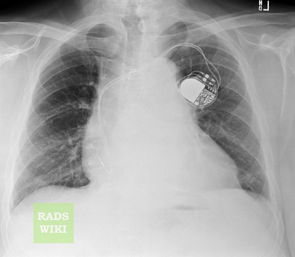
| [http://www.radswiki.net Images shown below are courtesy of RadsWiki]
| |
| | |
| [[Image:Pericard-effusion-01.jpg|thumb|350px|left|Pericardial effusion Day of the admission]]
| |
| <br clear="left"/>
| |
| | |
| [[Image:Pericard-effusion-02.jpg|thumb|350px|left|Pericardial effusion. The second day of treatment.]] | |
| <br clear="left"/>
| |
| | |
| ===Echocardiography===
| |
| | |
| [[Image:Hemorragic effusion.jpg|left|thumb|350px|A very large pericardial effusion due to malignancy as seen on cardiac ultrasound. Closed arrow: The heart, open arrow: The effusion]]
| |
| <br clear="left"/>
| |
| | |
| ===CT and MRI===
| |
| | |
| Cross-sectional imaging by CT or MRI is very sensitive in the detection of generalized or loculated pericardial effusions. Some fluid in the pericardial sac contributes to the apparent thickness and should be considered normal. Commonly, free-flowing fluid accumulates first at the posterolateral aspect of the left ventricle, when the patient is imaged in the supine position.
| |
| | |
| Estimation of the amount of fluid is possible to a limited extent based on the overall thickness of the crescent of fluid. Compared to cardiac ultrasound, CT and MRI may be particularly helpful in detecting loculated effusions, owing to the wide field of view provided by these techniques. Hemorrhagic effusions can be differentiated from a transudate or an exudate based on signal characteristics (high signal on T1-weighted images) or density (high-density clot on CT). Pulsation artefacts may cause local areas of low signal in a hemorrhagic effusion. Effusions are often incidentally noted on CT scans obtained for other indications.
| |
| | |
| Pericardial thickening (thickness >4 mm) is difficult to differentiate from a small generalized effusion. Both entities will reveal a low signal/density line that is thicker than the normal pericardial thickness. In acute pericarditis, the pericardial lining can show intermediate signal intensity and may enhance after gadolinium administration.
| |
| | |
| ====CT====
| |
| | |
| *CT attenuation measurements also enable the initial characterization of pericardial fluid.
| |
| *A fluid collection with attenuation close to that of water is likely to be a simple effusion.
| |
| *Attenuation greater than that of water suggests malignancy, hemopericardium, purulent exudate, or effusion associated with hypothyroidism.
| |
| *Pericardial effusions with low attenuation also have been reported in cases of chylopericardium.
| |
| | |
| [http://www.radswiki.net Images shown below are courtesy of RadsWiki]
| |
| | |
| [[Image:CT pericardial effusion.jpg|thumb|350px|left|CT: pericardial effusion]]
| |
| <br clear="left"/>
| |
| | |
| [[Image:Pericard-effusion-03.jpg|thumb|350px|left|Pericardial effusion. Second day of the admission.]]
| |
| <br clear="left"/>
| |
| | |
| ====MRI====
| |
| | |
| *The appearance of pericardial fluid is different on SE and GRE cine MR images.
| |
| *Nonhemorrhagic fluid has low signal intensity on T1-weighted SE images and high intensity on GRE cine images. Conversely, hemorrhagic effusion is characterized by high signal intensity on T1-weighted SE images and low intensity on GRE cine images.
| |
| *When an effusion is secondary to malignancy, an irregularly thickened pericardium or pericardial nodularity may be depicted on MR images.
| |
| | |
| ===Cardiac Catheterization===
| |
| | |
| Flouroscopic images show pericardial effusion:
| |
| | |
| <googlevideo>-7129815717409714366&hl=en</googlevideo>
| |
| | |
| | |
| <googlevideo>7051277599064845698&hl=en</googlevideo>
| |
| | |
| | |
| <googlevideo>1013614061451207857&hl=en</googlevideo>
| |
| | |
| | |
| <googlevideo>3444457597731375301&hl=en</googlevideo>
| |
| | |
| ==Treatment==
| |
| Treatment depends on the underlying cause and the severity of the [[heart impairment]]. Pericardial effusion due to a viral infection usually goes away within a few weeks without treatment. Some pericardial effusions remain small and never need treatment. If the pericardial effusion is due to a condition such as [[lupus]], treatment with anti-inflammatory medications may help. If the effusion is compromising heart function and causing cardiac [[tamponade]], it will need to be drained, most commonly by a needle inserted through the chest wall and into the pericardial space. A drainage tube is often left in place for several days. In some cases, surgical drainage may be required by [[pericardiocentesis]], in which a needle, and sometimes a [[catheter]] are used to drain excess fluid.
| |
| | |
| ==References==
| |
| <references />
| |
|
| |
|
| | ==See also== |
| | *[[Pericarditis]] |
| | *[[Constrictive pericarditis]] |
| | *[[Cardiac tamponade]] |
| <br> | | <br> |
|
| |
|
| [[es:Efusión pericárdica]] | | [[es:Efusión pericárdica]] |
|
| |
| {{Circulatory system pathology}}
| |
| {{Electrocardiography}}
| |
| {{SIB}}
| |
|
| |
|
| [[Category:Diseases involving the fasciae]] | | [[Category:Diseases involving the fasciae]] |
