Nonpathogenic intestinal amebae infection laboratory findings: Difference between revisions
Esther Lee (talk | contribs) No edit summary |
Esther Lee (talk | contribs) No edit summary |
||
| Line 10: | Line 10: | ||
====Endolimax nana==== | ====Endolimax nana==== | ||
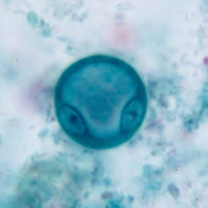
Cysts of Endolimax nana are spherical to ellipsoidal in shape and measure 5 to 10 µm. Mature cysts possess four nuclei with large, karyosomes and no peripheral chromatin. The nuclei are not visible in unstained wet mounts, but are visible in iodine-stained wet mounts and permanent slides stained with trichrome. The cytoplasm may contain diffuse glycogen, but lacks chromatoid bodies. | Cysts of Endolimax nana are spherical to ellipsoidal in shape and measure 5 to 10 µm. Mature cysts possess four nuclei with large, karyosomes and no peripheral chromatin. The nuclei are not visible in unstained wet mounts, but are visible in iodine-stained wet mounts and permanent slides stained with trichrome. The cytoplasm may contain diffuse glycogen, but lacks chromatoid bodies. | ||
---- | |||
*Shown below is a cyst of E. nana in a direct wet mount stained with iodine. | *Shown below is a cyst of E. nana in a direct wet mount stained with iodine. | ||
[[image:Enana_cyst_wtmt_lugols.jpg|Cyst of E. nana in a direct wet mount stained with iodine]] | [[image:Enana_cyst_wtmt_lugols.jpg|Cyst of E. nana in a direct wet mount stained with iodine]] | ||
---- | |||
*Shown below is a cyst of E. nana in a direct wet mount, viewed under differential interference contrast (DIC) microscopy. | *Shown below is a cyst of E. nana in a direct wet mount, viewed under differential interference contrast (DIC) microscopy. | ||
[[image:Enana_cyst_wtmt2.jpg|Cyst of E. nana in a direct wet mount, viewed under differential interference contrast (DIC) microscopy]] | [[image:Enana_cyst_wtmt2.jpg|Cyst of E. nana in a direct wet mount, viewed under differential interference contrast (DIC) microscopy]] | ||
---- | |||
*Shown below are cysts of E. nana stained with trichrome. | *Shown below are cysts of E. nana stained with trichrome. | ||
[[image:Enana_cyst_tric.jpg|Cysts of E. nana stained with trichrome]] | [[image:Enana_cyst_tric.jpg|Cysts of E. nana stained with trichrome]] | ||
[[image:E_nana_cyst_trich_BAM3.jpg|Cysts of E. nana stained with trichrome]] | [[image:E_nana_cyst_trich_BAM3.jpg|Cysts of E. nana stained with trichrome]] | ||
---- | |||
*Shown below are trophozoites of E. nana stained with trichrome. | *Shown below are trophozoites of E. nana stained with trichrome. | ||
[[image:Enana_troph_trich2.jpg|Trophozoites of E. nana stained with trichrome]] | [[image:Enana_troph_trich2.jpg|Trophozoites of E. nana stained with trichrome]] | ||
[[image:Enana_troph_tric3.jpg|Trophozoites of E. nana stained with trichrome]] | [[image:Enana_troph_tric3.jpg|Trophozoites of E. nana stained with trichrome]] | ||
---- | |||
====Entamoeba coli==== | ====Entamoeba coli==== | ||
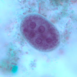
Cysts of Entamoeba coli are usually spherical, but may be elongated, and measure 10 to 35 µm. Mature cysts typically have 8 nuclei but may have as many as 16 or more. Entamoeba coli is the only species in the genus encountered in humans with more than four nuclei in the cyst stage. The nuclei may be seen in unstained as well as stained specimens. Karyosomes may be compact or diffuse, and are usually eccentrically located. Peripheral chromatin is present and is often coarse and granular, and irregularly arranged along the nuclear membrane, but may be more uniform. The cytoplasm of mature cysts may contain diffuse glycogen. Chromatoid bodies are seen less frequently than in E. histolytica. When present, they are usually splinter like with pointed ends and thus different from the chromatoid bodies of E. histolytica, which have rounded ends. | Cysts of Entamoeba coli are usually spherical, but may be elongated, and measure 10 to 35 µm. Mature cysts typically have 8 nuclei but may have as many as 16 or more. Entamoeba coli is the only species in the genus encountered in humans with more than four nuclei in the cyst stage. The nuclei may be seen in unstained as well as stained specimens. Karyosomes may be compact or diffuse, and are usually eccentrically located. Peripheral chromatin is present and is often coarse and granular, and irregularly arranged along the nuclear membrane, but may be more uniform. The cytoplasm of mature cysts may contain diffuse glycogen. Chromatoid bodies are seen less frequently than in E. histolytica. When present, they are usually splinter like with pointed ends and thus different from the chromatoid bodies of E. histolytica, which have rounded ends. | ||
---- | |||
*Shown below is a cyst of E. coli in a concentrated wet mount stained with iodine. Five nuclei are visible in this focal plane. | *Shown below is a cyst of E. coli in a concentrated wet mount stained with iodine. Five nuclei are visible in this focal plane. | ||
[[image:Ecoli_cyst_wtmt2.jpg|Cyst of E. coli in a concentrated wet mount stained with iodine. Five nuclei are visible in this focal plane]] | [[image:Ecoli_cyst_wtmt2.jpg|Cyst of E. coli in a concentrated wet mount stained with iodine. Five nuclei are visible in this focal plane]] | ||
---- | |||
*Shown bleow is a cyst of E. coli in a concentrated wet mount stained with iodine. Seven nuclei are visible in this focal plane. | *Shown bleow is a cyst of E. coli in a concentrated wet mount stained with iodine. Seven nuclei are visible in this focal plane. | ||
[[image:Ecoli_cyst_wtmt3.jpg|Cyst of E. coli in a concentrated wet mount stained with iodine. Seven nuclei are visible in this focal plane]] | [[image:Ecoli_cyst_wtmt3.jpg|Cyst of E. coli in a concentrated wet mount stained with iodine. Seven nuclei are visible in this focal plane]] | ||
---- | |||
*Shown below are the same cyst in two different focal planes. Eight nuclei can be seen between the two focal planes. Also, above the cyst in the first image, a trophozoite of Endolimax nana can be seen. | *Shown below are the same cyst in two different focal planes. Eight nuclei can be seen between the two focal planes. Also, above the cyst in the first image, a trophozoite of Endolimax nana can be seen. | ||
[[image:E_coli_cyst_BAM1a.jpg|Mature cyst of E. coli, stained with trichrome. These two images represent the same cyst shown in two different focal planes. Eight nuclei can be seen between the two focal planes. Also, above the cyst in the first image, a trophozoite of Endolimax nana can be seen]] | [[image:E_coli_cyst_BAM1a.jpg|Mature cyst of E. coli, stained with trichrome. These two images represent the same cyst shown in two different focal planes. Eight nuclei can be seen between the two focal planes. Also, above the cyst in the first image, a trophozoite of Endolimax nana can be seen]] | ||
[[image:E_coli_cyst_BAM1b.jpg|Mature cyst of E. coli, stained with trichrome. These two images represent the same cyst shown in two different focal planes. Eight nuclei can be seen between the two focal planes. Also, above the cyst in the first image, a trophozoite of Endolimax nana can be seen]] | [[image:E_coli_cyst_BAM1b.jpg|Mature cyst of E. coli, stained with trichrome. These two images represent the same cyst shown in two different focal planes. Eight nuclei can be seen between the two focal planes. Also, above the cyst in the first image, a trophozoite of Endolimax nana can be seen]] | ||
---- | |||
*Shown below are immature cyst of E. coli, stained with trichrome. Notice the presence of only two nuclei, and a large glycogen vacuole[[image:Ecoli_cyst_tric.jpg|Immature cyst of E. coli, stained with trichrome. Notice the presence of only two nuclei, and a large glycogen vacuole]] | *Shown below are immature cyst of E. coli, stained with trichrome. Notice the presence of only two nuclei, and a large glycogen vacuole[[image:Ecoli_cyst_tric.jpg|Immature cyst of E. coli, stained with trichrome. Notice the presence of only two nuclei, and a large glycogen vacuole]] | ||
---- | |||
*Shown below are mature mature cyst of E. coli, stained with trichrome. In this specimens, five nuclei are visible in the shown focal plane.[[image:Ecoli_cyst_tric3.jpg|Mature cyst of E. coli, stained with trichrome. In this specimens, five nuclei are visible in the shown focal plane]] | *Shown below are mature mature cyst of E. coli, stained with trichrome. In this specimens, five nuclei are visible in the shown focal plane.[[image:Ecoli_cyst_tric3.jpg|Mature cyst of E. coli, stained with trichrome. In this specimens, five nuclei are visible in the shown focal plane]] | ||
---- | |||
*Shown below are trophozoites of E. coli stained with trichrome. | *Shown below are trophozoites of E. coli stained with trichrome. | ||
[[image:Ecoli_troph_tric3.jpg|Trophozoites of E. coli stained with trichrome]] | [[image:Ecoli_troph_tric3.jpg|Trophozoites of E. coli stained with trichrome]] | ||
[[image:E_coli_troph_BAM1.jpg|Trophozoites of E. coli stained with trichrome]] | [[image:E_coli_troph_BAM1.jpg|Trophozoites of E. coli stained with trichrome]] | ||
---- | |||
==Sources== | ==Sources== | ||
*http://www.dpd.cdc.gov/dpdx/HTML/Frames/G-L/IntestinalAmebae/body_IntestinalAmebae_page2.htm#Laboratory%20Diagnosis | *http://www.dpd.cdc.gov/dpdx/HTML/Frames/G-L/IntestinalAmebae/body_IntestinalAmebae_page2.htm#Laboratory%20Diagnosis | ||
Revision as of 17:41, 19 November 2012
|
Nonpathogenic intestinal amebae infection Microchapters |
|
Differentiating Nonpathogenic intestinal amebae infection from other Diseases |
|---|
|
Diagnosis |
|
Treatment |
|
Case Studies |
|
Nonpathogenic intestinal amebae infection laboratory findings On the Web |
|
American Roentgen Ray Society Images of Nonpathogenic intestinal amebae infection laboratory findings |
|
FDA on Nonpathogenic intestinal amebae infection laboratory findings |
|
CDC on Nonpathogenic intestinal amebae infection laboratory findings |
|
Nonpathogenic intestinal amebae infection laboratory findings in the news |
|
Blogs on Nonpathogenic intestinal amebae infection laboratory findings |
|
Risk calculators and risk factors for Nonpathogenic intestinal amebae infection laboratory findings |
Editor-In-Chief: C. Michael Gibson, M.S., M.D. [1]
Overview
For E. coli, E. hartmanni, E. polecki, E. nana, and I. buetschlii, identification is made by observing cysts and/or trophozoites in stool specimens, both concentrated wet mounts and permanent stained smears.
Other Diagnostic Studies
Microscopy
Endolimax nana
Cysts of Endolimax nana are spherical to ellipsoidal in shape and measure 5 to 10 µm. Mature cysts possess four nuclei with large, karyosomes and no peripheral chromatin. The nuclei are not visible in unstained wet mounts, but are visible in iodine-stained wet mounts and permanent slides stained with trichrome. The cytoplasm may contain diffuse glycogen, but lacks chromatoid bodies.
- Shown below is a cyst of E. nana in a direct wet mount stained with iodine.

- Shown below is a cyst of E. nana in a direct wet mount, viewed under differential interference contrast (DIC) microscopy.

- Shown below are cysts of E. nana stained with trichrome.


- Shown below are trophozoites of E. nana stained with trichrome.


Entamoeba coli
Cysts of Entamoeba coli are usually spherical, but may be elongated, and measure 10 to 35 µm. Mature cysts typically have 8 nuclei but may have as many as 16 or more. Entamoeba coli is the only species in the genus encountered in humans with more than four nuclei in the cyst stage. The nuclei may be seen in unstained as well as stained specimens. Karyosomes may be compact or diffuse, and are usually eccentrically located. Peripheral chromatin is present and is often coarse and granular, and irregularly arranged along the nuclear membrane, but may be more uniform. The cytoplasm of mature cysts may contain diffuse glycogen. Chromatoid bodies are seen less frequently than in E. histolytica. When present, they are usually splinter like with pointed ends and thus different from the chromatoid bodies of E. histolytica, which have rounded ends.
- Shown below is a cyst of E. coli in a concentrated wet mount stained with iodine. Five nuclei are visible in this focal plane.

- Shown bleow is a cyst of E. coli in a concentrated wet mount stained with iodine. Seven nuclei are visible in this focal plane.

- Shown below are the same cyst in two different focal planes. Eight nuclei can be seen between the two focal planes. Also, above the cyst in the first image, a trophozoite of Endolimax nana can be seen.


- Shown below are immature cyst of E. coli, stained with trichrome. Notice the presence of only two nuclei, and a large glycogen vacuole

- Shown below are mature mature cyst of E. coli, stained with trichrome. In this specimens, five nuclei are visible in the shown focal plane.

- Shown below are trophozoites of E. coli stained with trichrome.

