Meningioma CT
|
Meningioma Microchapters |
|
Diagnosis |
|---|
|
Treatment |
|
Case Studies |
|
Meningioma CT On the Web |
|
American Roentgen Ray Society Images of Meningioma CT |
Editor-In-Chief: C. Michael Gibson, M.S., M.D. [1] Associate Editor(s)-in-Chief: Haytham Allaham, M.D. [2]
Overview
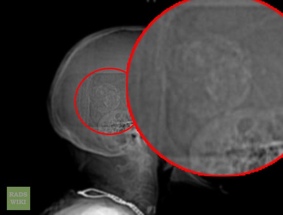
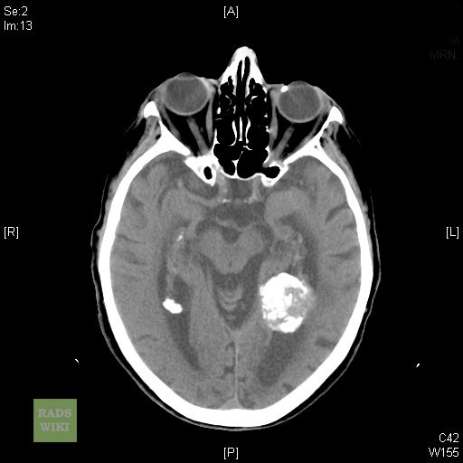
Head CT scan may be diagnostic of meningioma. Findings on CT scan suggestive of meningioma include homogeneously hyperdense lesion, calcification, hyperostosis, lytic lesions and pneumosinus dilatans.[1]
CT Scan
- Head CT scan may be diagnostic of meningioma.[1]
- Findings on CT scan suggestive of meningioma include:[1]
- Homogeneously hyperdense lesion
- Calcification
- Hyperostosis
- Lytic lesions
- Pneumosinus dilatans.
- Meningioma is highly vascularized thus it is readily visualized with contrast head CT scan.[1]
- Head CT scan confirms the location of the meningioma and illustrates any invasion to the surrounding brain tissue.
- Head CT scan may be used to determine if the meningioma could be surgically resected.
Gallery
Patient #1: 95 y/o female with a large intraventricular memingioma
-

CT scout
-
CT scout
-
-


Patient #2


Patient #3: Biopsy revealed atypical menigiomas


References
- ↑ 1.0 1.1 1.2 1.3 Meningioma. Radiopaedia(2015) http://radiopaedia.org/articles/meningioma Accessed on September, 25th 2015