Lymphangioma pathophysiology
|
Lymphangioma Microchapters |
|
Diagnosis |
|---|
|
Treatment |
|
Case Studies |
|
Lymphangioma pathophysiology On the Web |
|
American Roentgen Ray Society Images of Lymphangioma pathophysiology |
|
Risk calculators and risk factors for Lymphangioma pathophysiology |
Editor-In-Chief: C. Michael Gibson, M.S., M.D. [1] Associate Editor(s)-in-Chief: Haytham Allaham, M.D. [2]
Overview
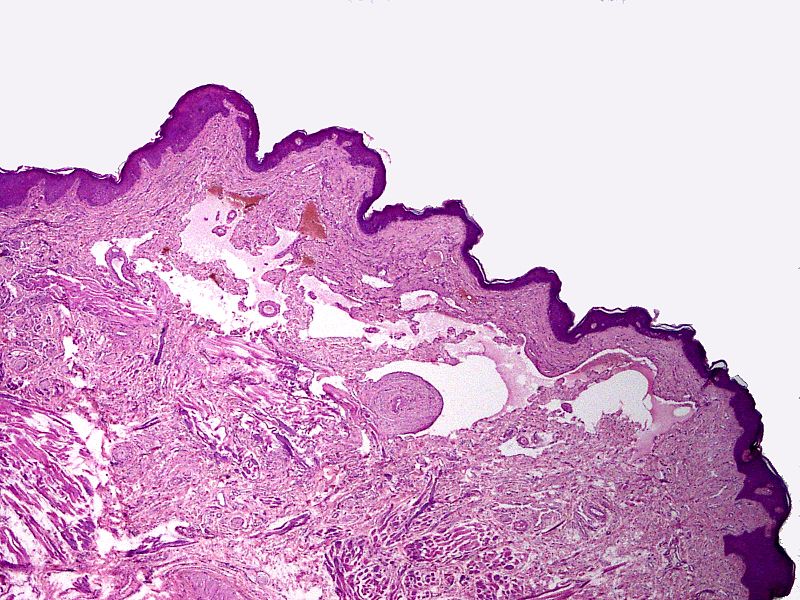
Lymphangioma arises from lymph vessels, which are normally involved in the re-circulation of excess body fluid back into the blood stream. The exact pathogenesis of lymphangioma is not fully understood. It is thought that lymphangioma is caused by either sequestration of lymph tissue, abnormal budding of lymph vessels, lack of fusion with the venous system, or obstruction of lymph vessels. Lymphangiomas most commonly develop at the head and neck regions. Lymphangioma is associated with a number of conditions that include Turner syndrome and Down syndrome. On gross pathology, characteristic findings of lymphangioma include a grey-white, well circumscribed, edematous mass with a variable size and consistency. On microscopic histopathological analysis, characteristic findings of lymphangioma include thin walled endothelial lining, intraluminal accumulation of eosinophilic deposits, and clusters of intraluminal lymphocytes.
Pathogenesis
- Lymphangioma arises from lymphatic vessels, which are normally involved in the re-circulation of excess body fluid back into the blood stream.
- Lymphangioma is a common benign tumor that often grows in proportion to the patients body growth rate.
- The exact pathogenesis of lymphangioma is not fully understood. It is thought that lymphangioma is caused by either sequestration of lymph tissue, abnormal budding of lymph vessels, lack of fusion with the venous system, or obstruction of lymph vessels.
- The progression to lymphangioma usually involves the following growth factors:
- VEGF-C
- VEGFR-3
- Prox-1
- bFGF
- PEDF
- Thrombospondin
- Reelin
- cMAF
- Integrin-α1
- Integrin-α9
- Lymphangiomas most commonly develop at the head and neck regions.
- However, lymphangioma may also develop in other anatomical sites such as:
- Breast
- Upper and lower limbs
- Internal visceral organs
Genetics
- Genetic mutations involved in the pathogenesis of lymphangioma include:
- Trisomy 13
- Tirsomy 18
- Trisomy 21
Associated Conditions
- Lymphangioma is associated with a number of conditions that include:
Gross Pathology
- On gross pathology, characteristic findings of lymphangioma include:
- Grey-white mass
- Well circumscribed
- Edematous appearance
- Variable size (may be massive)
- Filled with serous fluid
- Smooth inner lining
Microscopic Pathology
- On microscopic histopathological analysis, characteristic findings of lymphangioma include:
- Thin walled channels lined by endothelium
- Intraluminal accumulation of eosinophilic deposits
- Clusters of intraluminal lymphocytes
- On immunohistochemistry, characteristic findings of lymphangioma include:
- D2-40 +ve

