Lung cancer chest x ray: Difference between revisions
| Line 14: | Line 14: | ||
[[Image:Thorax pa peripheres Bronchialcarcinom li OF markiert.jpg|left|thumb|350px|Chest x-ray showing a cancerous tumor in the left lung]] | [[Image:Thorax pa peripheres Bronchialcarcinom li OF markiert.jpg|left|thumb|350px|Chest x-ray showing a cancerous tumor in the left lung]] | ||
<br clear="left"/> | <br clear="left"/> | ||
==A Clinical Example of Disease Progression== | |||
Images shown below are courtesy of Cafer Zorkun MD and copylefted | |||
<gallery> | |||
Image:Small Cell Lung CA 001.JPG|Chest x-ray: Small cell carcinoma of the lung. At the time of diagnosis. | |||
Image:Small Cell Lung CA 002.JPG|Chest x-ray: Small cell carcinoma of the lung. Five weeks later. | |||
Image:Small Cell Lung CA 003.JPG|Chest x-ray: Small cell carcinoma of the lung. Two months later. | |||
</gallery> | |||
==References== | ==References== | ||
Revision as of 19:45, 4 December 2011
|
Lung cancer Microchapters |
|
Diagnosis |
|---|
|
Treatment |
|
Case Studies |
|
Lung cancer chest x ray On the Web |
|
American Roentgen Ray Society Images of Lung cancer chest x ray |
|
Risk calculators and risk factors for Lung cancer chest x ray |
Editor-In-Chief: C. Michael Gibson, M.S., M.D. [1]; Associate Editor(s)-In-Chief: Kim-Son H. Nguyen, M.D., M.P.A., Beth Israel Deaconess Medical Center, Harvard Medical School, Boston MA, Cafer Zorkun, M.D., Ph.D. [2]
Overview
Performing a chest x-ray is the first step if a patient reports symptoms that may be suggestive of lung cancer. Often lung cancers are picked up on a routine chest X-ray in a person experiencing no symptoms.
X-ray findings
This may reveal an obvious mass, widening of the mediastinum (suggestive of spread to lymph nodes there), atelectasis (collapse), consolidation (pneumonia), or pleural effusion.
If there are no x-ray findings but the suspicion is high (such as a heavy smoker with blood-stained sputum), bronchoscopy and/or a CT scan may provide the necessary information. Bronchoscopy or CT-guided biopsy is often used to identify the tumor type.
The differential diagnosis for patients who present with abnormalities on chest x-ray includes lung cancer, as well as nonmalignant diseases. These include infectious causes such as tuberculosis or pneumonia, or inflammatory conditions such assarcoidosis. These diseases can result in mediastinal lymphadenopathy or lung nodules, and sometimes mimic lung cancers.

A Clinical Example of Disease Progression
Images shown below are courtesy of Cafer Zorkun MD and copylefted
-
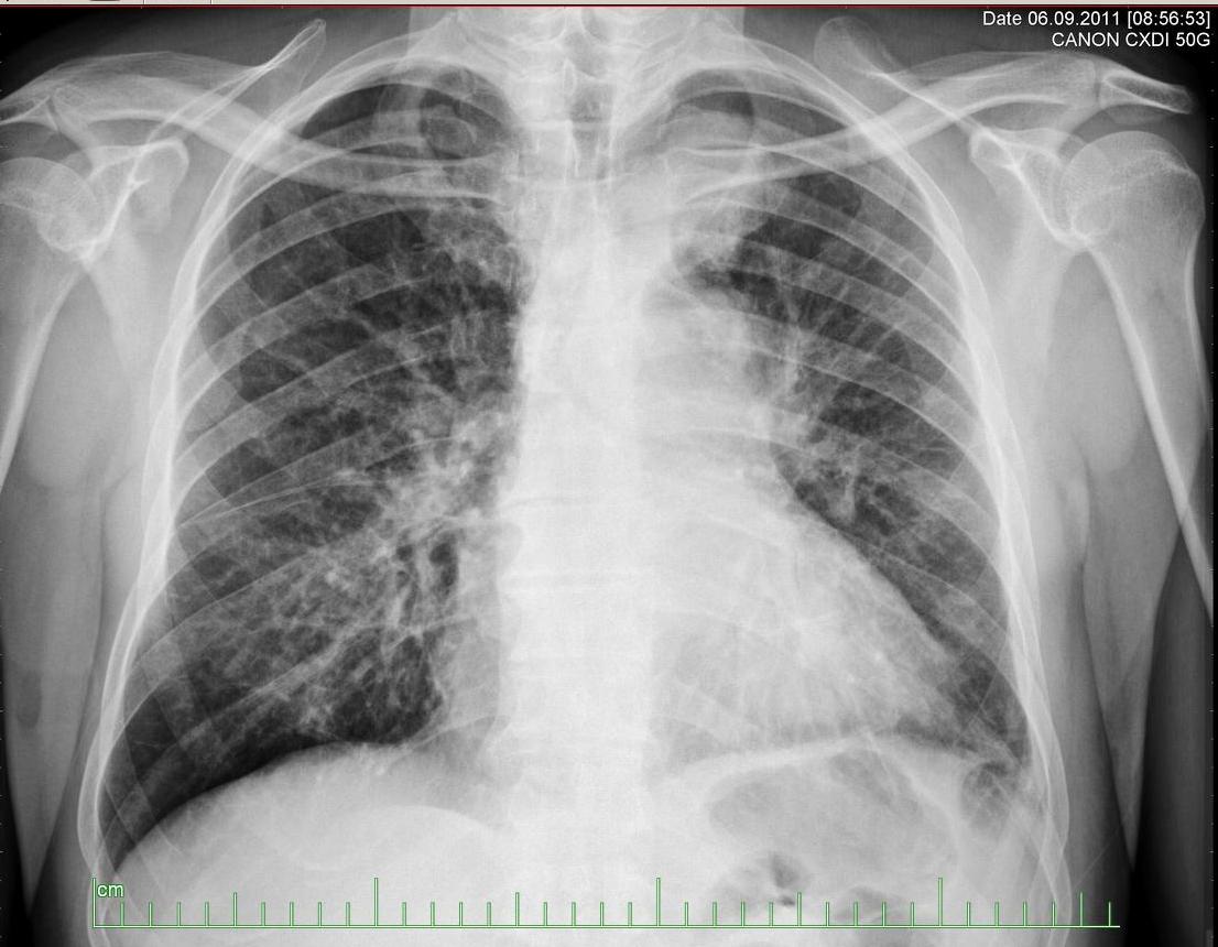
Chest x-ray: Small cell carcinoma of the lung. At the time of diagnosis.
-
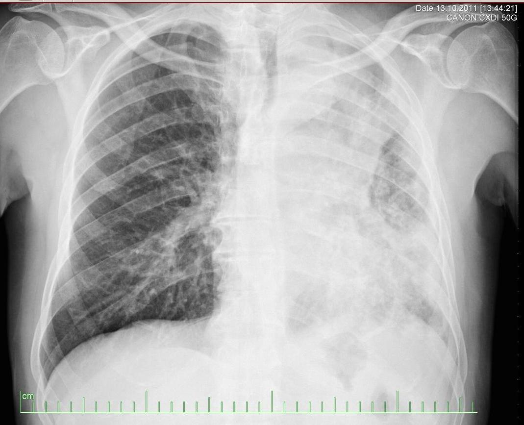
Chest x-ray: Small cell carcinoma of the lung. Five weeks later.
-
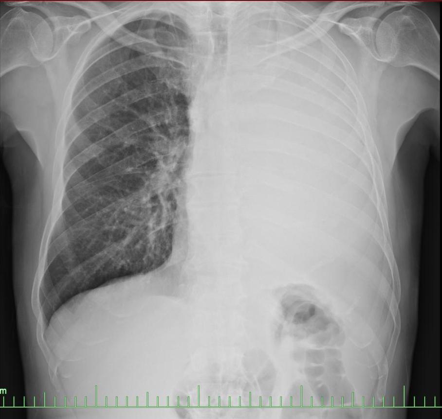
Chest x-ray: Small cell carcinoma of the lung. Two months later.


