Left atrial enlargement chest x-ray: Difference between revisions
Jump to navigation
Jump to search
Varun Kumar (talk | contribs) (Created page with "{{CMG}}; {{AOEIC}} {{VK}} ==Chest X-Ray== Chest x-ray findings of left atrial enlargement are: *Double density sign: Occur when the right side of the left atrium pushes behind...") |
Varun Kumar (talk | contribs) No edit summary |
||
| Line 1: | Line 1: | ||
{{CMG}}; {{AOEIC}} {{VK}} | {{CMG}}; {{AOEIC}} {{CZ}}; {{VK}} | ||
==Chest X-Ray== | ==Chest X-Ray== | ||
Chest x-ray findings of left atrial enlargement are: | Chest x-ray findings of left atrial enlargement are: | ||
| Line 8: | Line 8: | ||
*Superior displacement of the left main stem bronchus on frontal view | *Superior displacement of the left main stem bronchus on frontal view | ||
*Posterior displacement of a barium filled oesophagus or [[nasogastric tube]] | *Posterior displacement of a barium filled oesophagus or [[nasogastric tube]] | ||
Images shown below are courtesy of Radiopedia.com. | |||
<div align="left"> | |||
<gallery heights="175" widths="175"> | |||
Image:Left-atrial-enlargement-001.jpg|Double density sign | |||
Image:Left-atrial-enlargement-002.jpg|Same patient & the same image. Double density sign. Image's modified for more contrast and better visualization. | |||
Image:Left atrial enlargement with splaying of carina.jpg|Aside from the dirty lung due to [[emphysema]] and pneumonic infiltration in the lower right field you can notice a marked enlargement of the left atrium with splaying of the carina. | |||
</gallery> | |||
</div> | |||
==References== | ==References== | ||
Latest revision as of 14:23, 17 April 2012
Editor-In-Chief: C. Michael Gibson, M.S., M.D. [1]; Associate Editor(s)-In-Chief: Cafer Zorkun, M.D., Ph.D. [2]; Varun Kumar, M.B.B.S. [3]
Chest X-Ray
Chest x-ray findings of left atrial enlargement are:
- Double density sign: Occur when the right side of the left atrium pushes behind the right atrial border, appearing as a double density. If large enough it can actually reach beyond the border of the right atrium.
- Convex left atria appendage: usually reflect prior rheumatic heart disease
- Splaying of the carina
- Posterior displacement of the left main stem bronchus on lateral radiograph
- Superior displacement of the left main stem bronchus on frontal view
- Posterior displacement of a barium filled oesophagus or nasogastric tube
Images shown below are courtesy of Radiopedia.com.
-
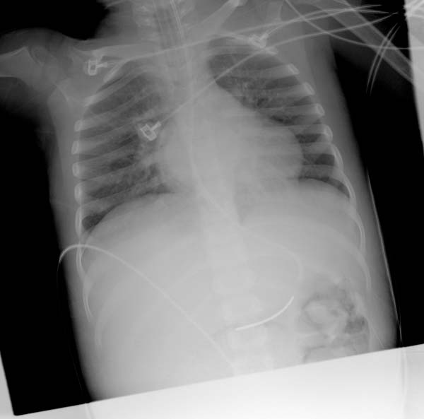
Double density sign
-
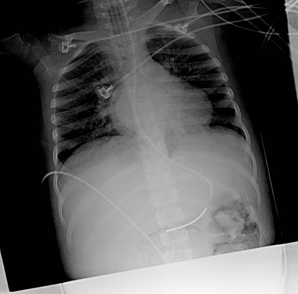
Same patient & the same image. Double density sign. Image's modified for more contrast and better visualization.
-
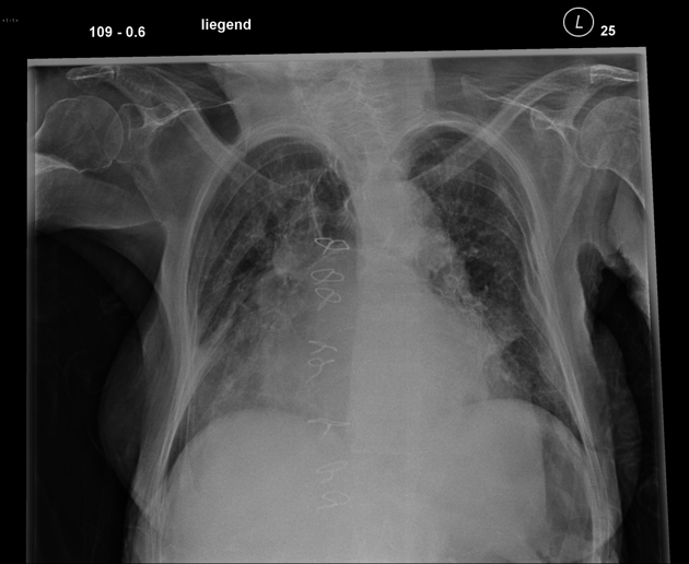
Aside from the dirty lung due to emphysema and pneumonic infiltration in the lower right field you can notice a marked enlargement of the left atrium with splaying of the carina.


