Laryngeal cancer pathophysiology
|
Laryngeal cancer Microchapters |
|
Diagnosis |
|---|
|
Treatment |
|
Case Studies |
|
Laryngeal cancer pathophysiology On the Web |
|
American Roentgen Ray Society Images of Laryngeal cancer pathophysiology |
|
Risk calculators and risk factors for Laryngeal cancer pathophysiology |
Editor-In-Chief: C. Michael Gibson, M.S., M.D. [1] Associate Editor(s)-in-Chief: Faizan Sheraz, M.D. [2]
Overview
Pathophysiology
Gross Pathology

Subclassification by site
It is generally divided the following way:[1]
| Laryngeal cancer | |||||||||||||||||||||||||||||||||
| Supraglottis | Glottis | Subglottis | |||||||||||||||||||||||||||||||
- Prevalence - glottis > supraglottis > subglottis.
- Glottic carcinoma tends to present earlier (as it affects phonation) and, therefore, has a better prognosis.
SCC is subdivided by the WHO into:[4]
- Keratinizing type (KT).
- Worst prognosis.
- Undifferentiated type (UT).
- Intermediate prognosis.
- EBV association.
- Nonkeratinizing type (NT).
- Good prognosis.
- EBV association.
Microscopic
Features based on classification:[4]
- KT subtype:
- Keratinization & intercellular bridges through-out most of the malignant lesion.
- UT:
- Non-distinct borders/syncytial pattern.
- Nucleoli.
- NT:
- Well-defined cell borders.
Invasion
Features:
- Eosinophilia.
- Extra large nuclei/bizarre nuclei.
- Inflammation (lymphocytes, plasma cells).
- Long rete ridges.
- Numerous beeds/blobs of epithelial cells that seem unlikely to be rete ridges.
Pitfalls:
- Tangential cuts.
- If you can trace the squamous cells from a gland to the surface it is less likely to be invasive cancer.
Notes on invasion:
- Nice review paper by Wenig.[5]
- See SCC of the cervix versus CIN III.
Images
-
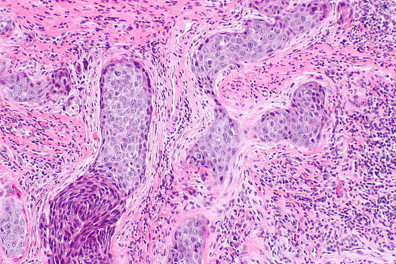
Laryngeal squamous carcinoma (Intermediate Magnification)
-
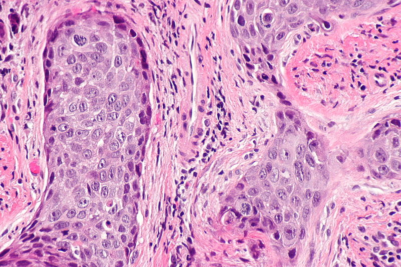
Laryngeal squamous carcinoma (High Magnification)
-
Laryngeal SCC - very high mag.


Overview of subtypes
There are several subtypes:[6]
- Basaloid - poor prognosis, usu. diagnosed by recognition of typical SCC.
- Warty (Condylomatous).
- Verrucous - good prognosis, rare.
- Papillary.
- Lymphoepithelial, rare.
- Spindle cell, a common spindle cell lesion of the H&N.
Verrucous squamous cell carcinoma
Features:
- Exophytic growth.
- Well-differentiated.
- "Glassy" appearance.
- Pushing border.
DDx: papilloma.
Spindle cell squamous carcinoma
- Key to diagnosis is finding a component of conventional squamous cell carcinoma.
IHC:
- Typically keratin -ve.
- p63 +ve.
DDx:
- Spindle cell melanoma.
- Mesenchymal neoplasm.
Basaloid squamous cell carcinoma
- May mimic adenoid cystic carcinoma.
- Classically base of tongue.[7]
- Typically poor prognosis.
Features:
- Need keratinization. (???)
DDx:
- Neuroendocrine tumour.
Lymphoepithelial (squamous cell) carcinoma
IHC
- p63 +ve.
- EBER -ve.
- Positive suggests nasopharyngeal carcinoma.
- p16 -ve.
- Positive suggests HPV-associated head and neck squamous cell carcinoma.
- Bcl2 +ve/-ve.
- Positive = poor prognosis.[8]
References
- ↑ URL: http://www.cap.org/apps/docs/committees/cancer/cancer_protocols/2011/Larynx_11protocol.pdf. Accessed on: 2 May 2012.
- ↑ Template:Ref WMSP
- ↑ URL: http://www.health.am/cr/more/statistics-and-prognosis-for-cancer-of-the-larynx/. Accessed on: 2 May 2012.
- ↑ 4.0 4.1 Template:Ref Sternberg4
- ↑ Wenig BM (2002). "Squamous cell carcinoma of the upper aerodigestive tract: precursors and problematic variants" (PDF). Mod. Pathol. 15 (3): 229–54. doi:10.1038/modpathol.3880520. PMID 11904340. Unknown parameter
|month=ignored (help) - ↑ URL: http://www.pathconsultddx.com/pathCon/diagnosis?pii=S1559-8675%2806%2970297-2. Accessed on: March 9, 2010.
- ↑ URL: http://www.biomedcentral.com/1471-2407/6/146. Accessed on: March 9, 2010.
- ↑ Nichols AC, Finkelstein DM, Faquin WC; et al. (2010). "Bcl2 and human papilloma virus 16 as predictors of outcome following concurrent chemoradiation for advanced oropharyngeal cancer". Clin. Cancer Res. 16 (7): 2138–46. doi:10.1158/1078-0432.CCR-09-3185. PMID 20233885. Unknown parameter
|month=ignored (help)