Hypertrophic cardiomyopathy pathologic abnormalities: Difference between revisions
(New page: {{Hypertrophic cardiomyopathy}} ==Overview== ==Gross Pathological Findings== <div align="left"> <gallery heights="175" widths="175"> Image:2367.jpg|Cardiomyopathy: Intermediate between h...) |
|||
| Line 35: | Line 35: | ||
</div> | </div> | ||
==References== | ==References== | ||
Latest revision as of 18:26, 14 August 2011
|
Hypertrophic Cardiomyopathy Microchapters |
|
Differentiating Hypertrophic Cardiomyopathy from other Diseases |
|---|
|
Diagnosis |
|
Treatment |
|
Case Studies |
|
Hypertrophic cardiomyopathy pathologic abnormalities On the Web |
|
Hypertrophic cardiomyopathy pathologic abnormalities in the news |
|
Blogs on Hypertrophic cardiomyopathy pathologic abnormalities |
|
Directions to Hospitals Treating Hypertrophic cardiomyopathy |
|
Risk calculators and risk factors for Hypertrophic cardiomyopathy pathologic abnormalities |
Overview
Gross Pathological Findings
-
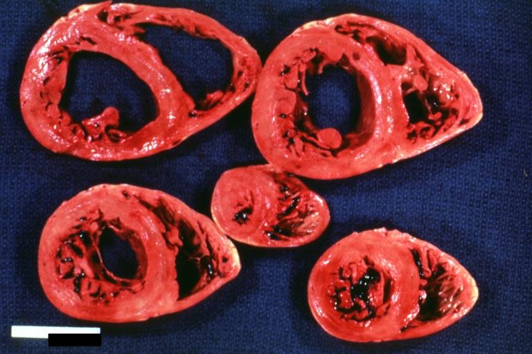
Cardiomyopathy: Intermediate between hypertrophic and dilated
-
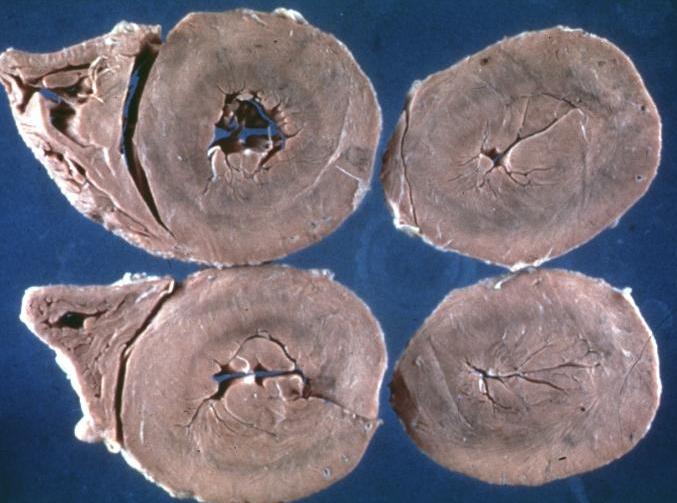
Hypertrophic cardiomyopathy, concentric


-
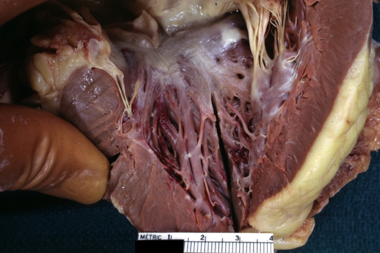
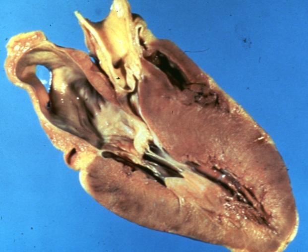
Hypertrophic Obstructive Cardiomyopathy: Gross natural color opened left ventricular outflow tract with subaortic shelf and marked endocardial thickening matching the contour of the anterior mitral leaflet
-
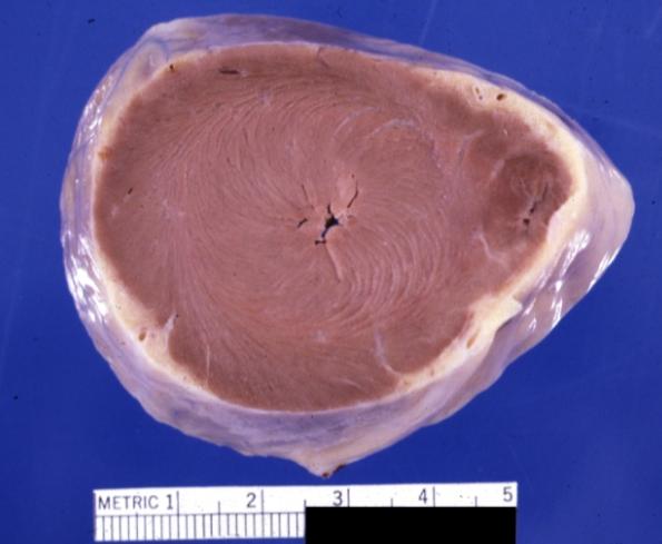
Cardiomyopathy: Gross apical slice of left and right ventricles concentric hypertrophy with cavitary obliteration sudden unexpected death obstructive cardiomyopathy


-
Cardiomyopathy Asymmetrical Septal Hypertrophy
-
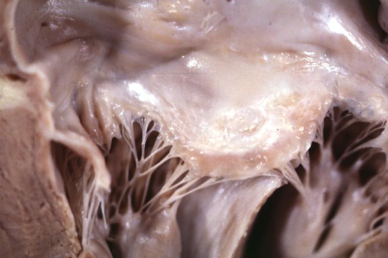
Cardiomyopathy: Gross excellent view of mitral valve atrial surface showing thickening which is fibrous in body of valve and myxoid at area of free margin changes presumed secondary to insufficiency due to anterior motion


-
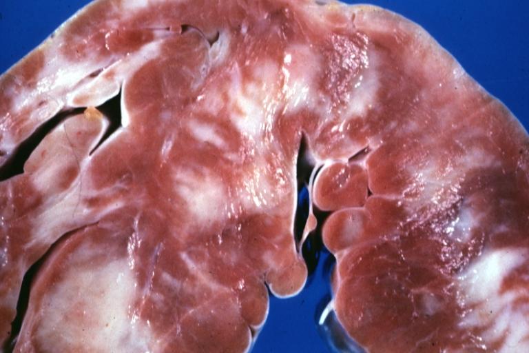
Cardiomyopathy: Gross close up view of a ventricle slice
-
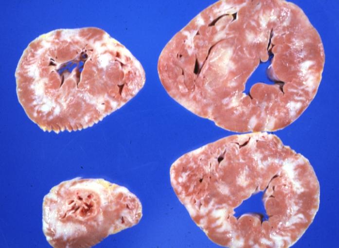
Cardiomyopathy: Gross ventricular slices hypertrophy and extensive myocardial fibrosis a unique case of global fiber disarray with atrophy and fibrosis

