Gallstone disease
Template:DiseaseDisorder infobox
|
Gallstone disease Microchapters |
|
Diagnosis |
|---|
|
Treatment |
|
Surgery |
|
Case Studies |
|
Gallstone disease On the Web |
|
American Roentgen Ray Society Images of Gallstone disease |
For patient information click here
Editor-In-Chief: C. Michael Gibson, M.S., M.D. [1]; Associate Editor-In-Chief: Cafer Zorkun, M.D., Ph.D. [2] Prashanth Saddala M.B.B.S
Synonyms and related keywords: Cholecystolithiasis; choleliths; cholelithiasis; biliary colic; gall stones; gallbladder calculus.
Overview
Historical Perspective
Classification
Pathophysiology
Causes
Differentiating Gallstone disease from other Diseases
Epidemiology and Demographics
Risk Factors
Screening
Natural History, Complications and Prognosis
Diagnosis
History and Symptoms | Physical Examination | Laboratory Findings | X Ray | CT | MRI | Echocardiography or Ultrasound | Other Imaging Findings | Other Diagnostic Studies
Treatment
Medical Therapy | Surgery | Primary Prevention | Secondary Prevention | Cost-Effectiveness of Therapy | Future or Investigational Therapies
Case Studies
Other Imaging Findings
- Endoscopic Retrograde Cholangiopancreatography (ERCP): most sensitive/specific for common bile duct (CBD) stones
- Magnetic Resonance Cholangiopancreatography (MRCP): diagnostic accuracy equivalent to ERCP, but not therapeutic
- Hepatobiliary Iminodiacetic Acid (HIDA) scan: highly sensitive for acute cholecytitis
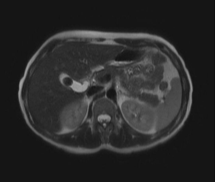
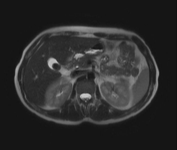
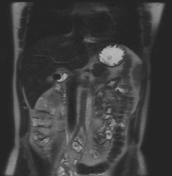
Patient #1: Gallstone on MRI
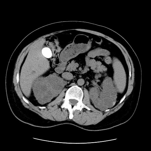
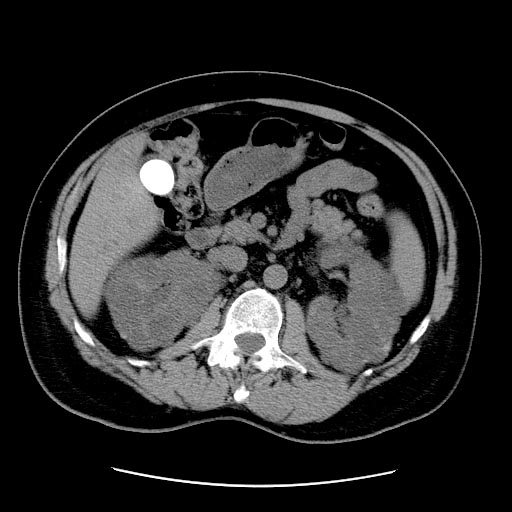
Patient #2: A large gallstone in a patient with Autosomal dominant polycystic kidney disease
Treatment
Nonoperative management is suboptimal (ursodiol, lithotripsy). Cholecystectomy is the therapy of choice.
Medical therapy
Cholesterol gallstones can sometimes be dissolved by oral ursodeoxycholic acid. Gallstones may recur however, once the drug is stopped. Obstruction of the common bile duct with gallstones can sometimes be relieved by endoscopic retrograde sphinceterotomy (ERS) following endoscopic retrograde cholangiopancreatography (ERCP). A common misconception is that the use of ultrasound (Extracorporeal Shock Wave Lithotripsy) can be used to break up gallstones. Although this treatment is highly effective against kidney stones, it can only rarely be used to break up the softer and less brittle gallstones.
Surgery
Cholecystectomy (gallbladder removal) has a 99% chance of eliminating the recurrence of cholelithiasis. Only symptomatic patients must be indicated to surgery. The lack of a gall bladder does not seem to have any negative consequences in many people. However, there is a significant proportion of the population, between 5-40%, who develop a condition called postcholecystectomy syndrome.[1] Symptoms include gastrointestinal distress and persistent pain in the upper right abdomen.
There are two surgery options: open procedure and laparoscopic: see the cholecystectomy article for more details.
- Open cholecystectomy procedure: This involves a large incision into the abdomen (laparotomy) below the right lower ribs. A week of hospitalization, normal diet a week after release and normal activity a month after release.
- Laparoscopic cholecystectomy: 3-4 small puncture holes for camera and instruments (available since the 1980s). Typically same-day release or one night hospital stay, followed by a week of home rest and pain medication. Can resume normal diet and light activity a week after release. (Decreased energy level and minor residual pain for a month or two.) Studies have shown that this procedure is as effective as the more invasive open cholecystectomy, provided the stones are accurately located by cholangiogram prior to the procedure so that they can all be removed. The procedure also has the benefit of reducing operative complications such as bowel perforation and vascular injury.
Alternative medicine
A regimen called a "gallbladder flush" or "liver flush" is a popular remedy in alternative medicine. In this treatment, often self-administered, the patient drinks four glasses of apple cider and eats five apples per day for five days, then fasts briefly, takes magnesium, and then drinks large quantities of lemon or grapefruit juice mixed with olive oil or other oil before bed; the next morning, they painlessly pass a number of green and brown pebbles purported to be stones flushed from the biliary system. A New Zealand hospital analyzed stones from a typical gallbladder flush and found them to be composed of fatty acids similar to those in olive oil, with no detectable cholesterol or bile salts,[2] demonstrating that they are little more than hardened olive oil. Despite the gallbladder flush, the patient still required surgical removal of multiple true gallstones. The note concluded: "The gallbladder flush may not be entirely worthless, however; there is one case report in which treatment with olive oil and lemon juice resulted in the passage of numerous gallstones, as demonstrated by ultrasound examination."[3]
In the case mentioned, ultrasound confirmed multiple gallstones, but after waiting months for a surgical option, the patient underwent a treatment with olive oil and lemon juice resulting in the passage of four 2.5 cm by 1.25 cm stones and twenty pea-sized stones. Two years later symptoms returned, and ultrasound showed a single large gallstone; the patient chose to have this removed surgically.[3]
References
- ↑ "Postcholecystectomy syndrome". WebMD. Retrieved 2007-08-25.
- ↑ Alan R. Gaby. "The gallstone cure that wasn't". Townsend Letter for Doctors and Patients. Retrieved 2007-02-10.
- ↑ 3.0 3.1 A. P. Savage (1992). "Case report. Adjuvant herbal treatment for gallstones". British Journal of Surgery. 79 (2): 168. Unknown parameter
|month=ignored (help); Unknown parameter|coauthors=ignored (help);|access-date=requires|url=(help)
External links
- Gallbladder removal - series from Nuggets Medical Encyclopedia
- "Gall Stones and Gall Bladder Surgery" Information for this article was located from the University of Florida, Medical Library in Gainesville, Florida, and researched by Richard Pressinger (M.Ed.) and Jerry Abraham (M.B.A)
- Diagram of pain radiation
