File:Epidemic typhus07.jpeg
Epidemic_typhus07.jpeg (700 × 475 pixels, file size: 51 KB, MIME type: image/jpeg)
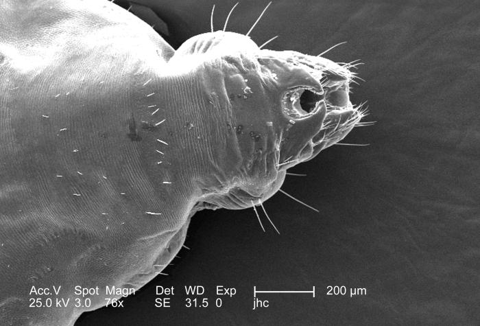
At double the magnification of PHIL, this 2006 scanning electron micrograph (SEM), magnified 152x, revealed the distal tip of the abdominal region of a female body louse, Pediculus humanus var. corporis from a dorsal perspective. Some of the morphologic characteristics seen in this image include the two “gonopodia”, which are located dorsal to the larger two setae-bearing “claspers”. It is into this notch that the male would insert the “aedeagus”, or penis during the process of copulation. This notch, identifying the louse as a female is observable to the naked eye, whereas, in the male louse, the distal abdomen is rounded, and not concave.
File history
Click on a date/time to view the file as it appeared at that time.
| Date/Time | Thumbnail | Dimensions | User | Comment | |
|---|---|---|---|---|---|
| current | 20:52, 8 December 2014 | | 700 × 475 (51 KB) | Jesus Hernandez (talk | contribs) | At double the magnification of PHIL, this 2006 scanning electron micrograph (SEM), magnified 152x, revealed the distal tip of the abdominal region of a female body louse, Pediculus humanus var. corporis from a dorsal perspective. Some of the morphologi... |
You cannot overwrite this file.
File usage
The following page uses this file: