File:Epidemic typhus06.jpeg
Epidemic_typhus06.jpeg (700 × 475 pixels, file size: 45 KB, MIME type: image/jpeg)
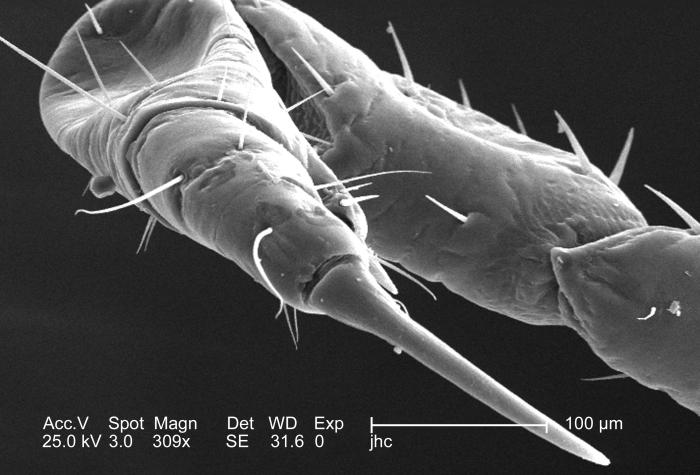
At a moderate magnification of 309x, this 2006 scanning electron micrograph (SEM) depicted an enlarged dorsal view of the right flexed foreleg of a female body louse, Pediculus humanus var. corporis. The entire leg is not quite visible, but what is visible includes the most distal segment, known as the “pretarsus”, followed by the more proximal “tarsus”, then the “tibia”, “femur”, and “trochanter”. The final segment, of the six segments from which each leg is composed, the “coxa”, is visible under reduced magnification, in PHIL# 9242. In the case of the louse, the leg segments are very stout, and end in claws, which it used to firmly grasp clothing, or a host’s hair shafts.
File history
Click on a date/time to view the file as it appeared at that time.
| Date/Time | Thumbnail | Dimensions | User | Comment | |
|---|---|---|---|---|---|
| current | 20:51, 8 December 2014 | | 700 × 475 (45 KB) | Jesus Hernandez (talk | contribs) | At a moderate magnification of 309x, this 2006 scanning electron micrograph (SEM) depicted an enlarged dorsal view of the right flexed foreleg of a female body louse, Pediculus humanus var. corporis. The entire leg is not quite visible, but what is vis... |
You cannot overwrite this file.
File usage
The following page uses this file: