File:Epidemic typhus03.jpeg
Epidemic_typhus03.jpeg (700 × 475 pixels, file size: 54 KB, MIME type: image/jpeg)
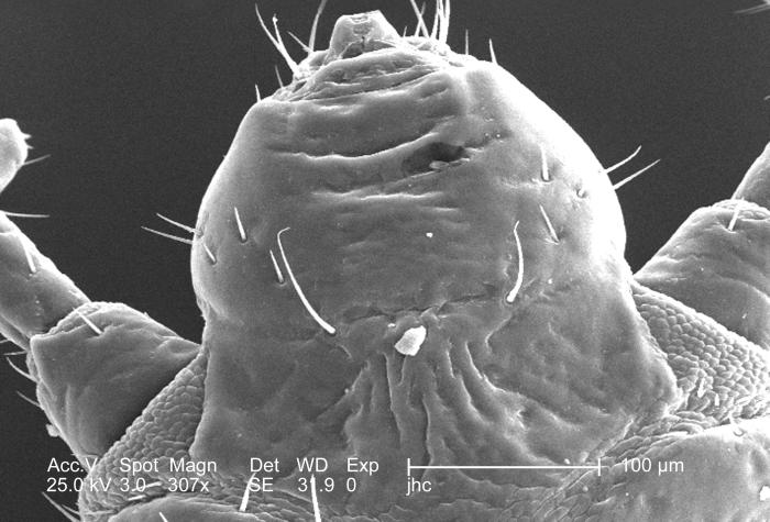
This was one of five scanning electron micrographic (SEM) images (PHIL# 9243 – 9247), successively magnified at higher and higher values, which focused on the head region of a female body louse, Pediculus humanus var. corporis from a ventral perspective. At a moderate magnification of 307x, this SEM revealed some of the insect’s exoskeletal morphology exhibited by the cephalic region. Note the two partially visible, bilaterally situated antennae composed of three main segments: visible here was the most proximal scape and the pedicle, and not in the field of view, the multi-segmented flagellum. The antennae, and the insect’s body sport sensorial “hairs” known as "setae”, both of which provided the organism with a "picture” of its environment, by taking readings in thermal, chemical, and mechanical changes encountered in its immediate surroundings. Its cone-shaped mouth is located at the very top of the image.
File history
Click on a date/time to view the file as it appeared at that time.
| Date/Time | Thumbnail | Dimensions | User | Comment | |
|---|---|---|---|---|---|
| current | 20:49, 8 December 2014 | | 700 × 475 (54 KB) | Jesus Hernandez (talk | contribs) | This was one of five scanning electron micrographic (SEM) images (PHIL# 9243 – 9247), successively magnified at higher and higher values, which focused on the head region of a female body louse, Pediculus humanus var. corporis from a ventral perspect... |
You cannot overwrite this file.
File usage
The following page uses this file: