File:Dracunculiasis07.jpeg
Dracunculiasis07.jpeg (700 × 525 pixels, file size: 47 KB, MIME type: image/jpeg)
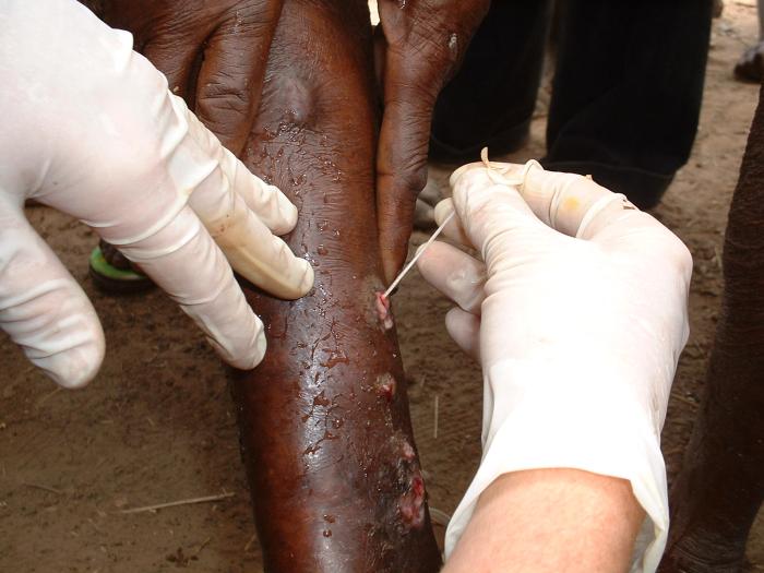
This image depicts the subcutaneous emergence of a female Guinea worm, Dracunculus medinensis, from a sufferer’s lower leg. Note the adjacent infected ulcerations surrounding the rupture site, representing other sites of worm emergence. Before the worm emerges, a blister develops on the skin. This blister causes a very painful burning sensation and eventually, ruptures within 24 - 72 hours. For relief, people will immerse the affected limb into water, whereupon, the adult female worm releases a milky white liquid containing millions of immature larvae into the water, thereby, contaminating the water supply. For several days after its emergence from the ulcer, the female Guinea worm is capable of releasing more larvae whenever it comes into contact with water.
File history
Click on a date/time to view the file as it appeared at that time.
| Date/Time | Thumbnail | Dimensions | User | Comment | |
|---|---|---|---|---|---|
| current | 14:59, 8 December 2014 | | 700 × 525 (47 KB) | Jesus Hernandez (talk | contribs) | This image depicts the subcutaneous emergence of a female Guinea worm, Dracunculus medinensis, from a sufferer’s lower leg. Note the adjacent infected ulcerations surrounding the rupture site, representing other sites of worm emergence. Before the wo... |
You cannot overwrite this file.
File usage
The following page uses this file: