File:Cryptosporidiosis04.jpeg
Original file (700 × 714 pixels, file size: 49 KB, MIME type: image/jpeg)
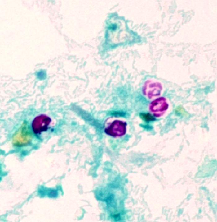
This photomicrograph revealed the morphologic details of Cryptosporidium parvum oocysts, i.e.,encapsulated zygotes, which had been stained using the modified acid-fast method. These oocysts exhibit a bright red coloration when using this staining technique, and in this case, you’ll note the sporozoites that were made visible inside the two oocysts on the right. Sporozoites are the nucleated, motile stage of development through which many protozoans pass such as C. parvum, on their way to becoming adults, and represent a very infectious form of these organisms. The sporozoites will be released from these C. parvum oocysts.
File history
Click on a date/time to view the file as it appeared at that time.
| Date/Time | Thumbnail | Dimensions | User | Comment | |
|---|---|---|---|---|---|
| current | 12:34, 5 December 2014 | | 700 × 714 (49 KB) | Jesus Hernandez (talk | contribs) | This photomicrograph revealed the morphologic details of Cryptosporidium parvum oocysts, i.e.,encapsulated zygotes, which had been stained using the modified acid-fast method. These oocysts exhibit a bright red coloration when using this staining techn... |
You cannot overwrite this file.
File usage
The following page uses this file: