File:Chromoblastomycosis04.jpeg
No higher resolution available.
Chromoblastomycosis04.jpeg (700 × 463 pixels, file size: 17 KB, MIME type: image/jpeg)
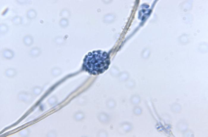
Magnified 1125X, this photomicrograph revealed some of the ultrastructural morphology exhibited by a cluster of Phialophora parasitica fungal conidia, which is attached to a conidiophore from which the former structure was derived.
File history
Click on a date/time to view the file as it appeared at that time.
| Date/Time | Thumbnail | Dimensions | User | Comment | |
|---|---|---|---|---|---|
| current | 20:50, 3 December 2014 | | 700 × 463 (17 KB) | Jesus Hernandez (talk | contribs) | Magnified 1125X, this photomicrograph revealed some of the ultrastructural morphology exhibited by a cluster of Phialophora parasitica fungal conidia, which is attached to a conidiophore from which the former structure was derived. |
You cannot overwrite this file.
File usage
The following page uses this file: