Failure to thrive CT scan

Revision as of 22:55, 13 September 2020 by Akash Daswaney (talk | contribs)
(diff) ← Older revision | Latest revision (diff) | Newer revision → (diff)
Jump to navigation Jump to search

Failure to thrive Microchapters

Home

Patient Information

Overview

Historical Perspective

Classification

Pathophysiology

Causes

Differentiating Failure to thrive from other Diseases

Epidemiology and Demographics

Risk Factors

Screening

Natural History, Complications and Prognosis

Diagnosis

Diagnostic Study of Choice

History and Symptoms

Physical Examination

Laboratory Findings

Electrocardiogram

X-ray

Echocardiography and Ultrasound

CT scan

MRI

Other Imaging Findings

Other Diagnostic Studies

Treatment

Medical Therapy

Interventions

Surgery

Primary Prevention

Secondary Prevention

Cost-Effectiveness of Therapy

Future or Investigational Therapies

Case Studies

Case #1

Failure to thrive CT scan On the Web

Most recent articles

Most cited articles

Review articles

CME Programs

Powerpoint slides

Images

American Roentgen Ray Society Images of Failure to thrive CT scan

All Images
X-rays
Echo & Ultrasound
CT Images
MRI

Ongoing Trials at Clinical Trials.gov

US National Guidelines Clearinghouse

NICE Guidance

FDA on Failure to thrive CT scan

CDC on Failure to thrive CT scan

Failure to thrive CT scan in the news

Blogs on Failure to thrive CT scan

Directions to Hospitals Treating Psoriasis

Risk calculators and risk factors for Failure to thrive CT scan

Editor-In-Chief: C. Michael Gibson, M.S., M.D. [1]; Associate Editor(s)-in-Chief: Akash Daswaney, M.B.B.S[2]

Overview

CTs are useful in diagnosing organic causes of failure to thrive.

CT scan

  • CTs are useful in diagnosing organic causes of failure to thrive.
  • Listing down each organic cause is beyond the scope of this microchapter.
  • Some examples are
  • Unilateral renal ectopia
    1. CT pelvis of a boy who presented failure to thrive, fatigue, intermittent fevers, weight loss, night sweats, and vague discomfort in his abdomen and extremities.
    2. CT shows the right kidney ectopically positioned in the right lower quadrant.
https://medpix.nlm.nih.gov/case?id=ed1c24d6-a7c5-402f-8f04-d0816b846c3a
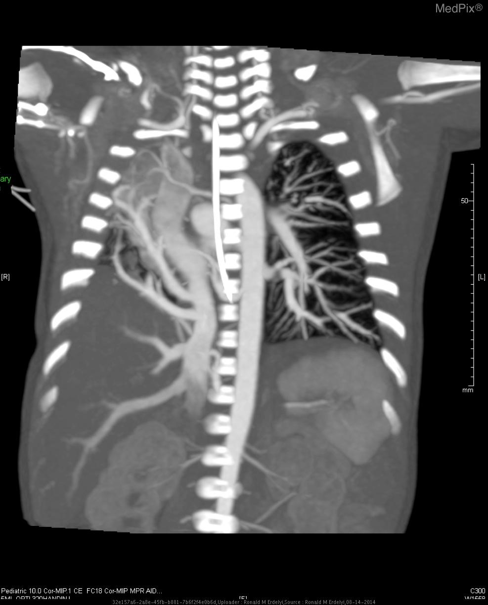
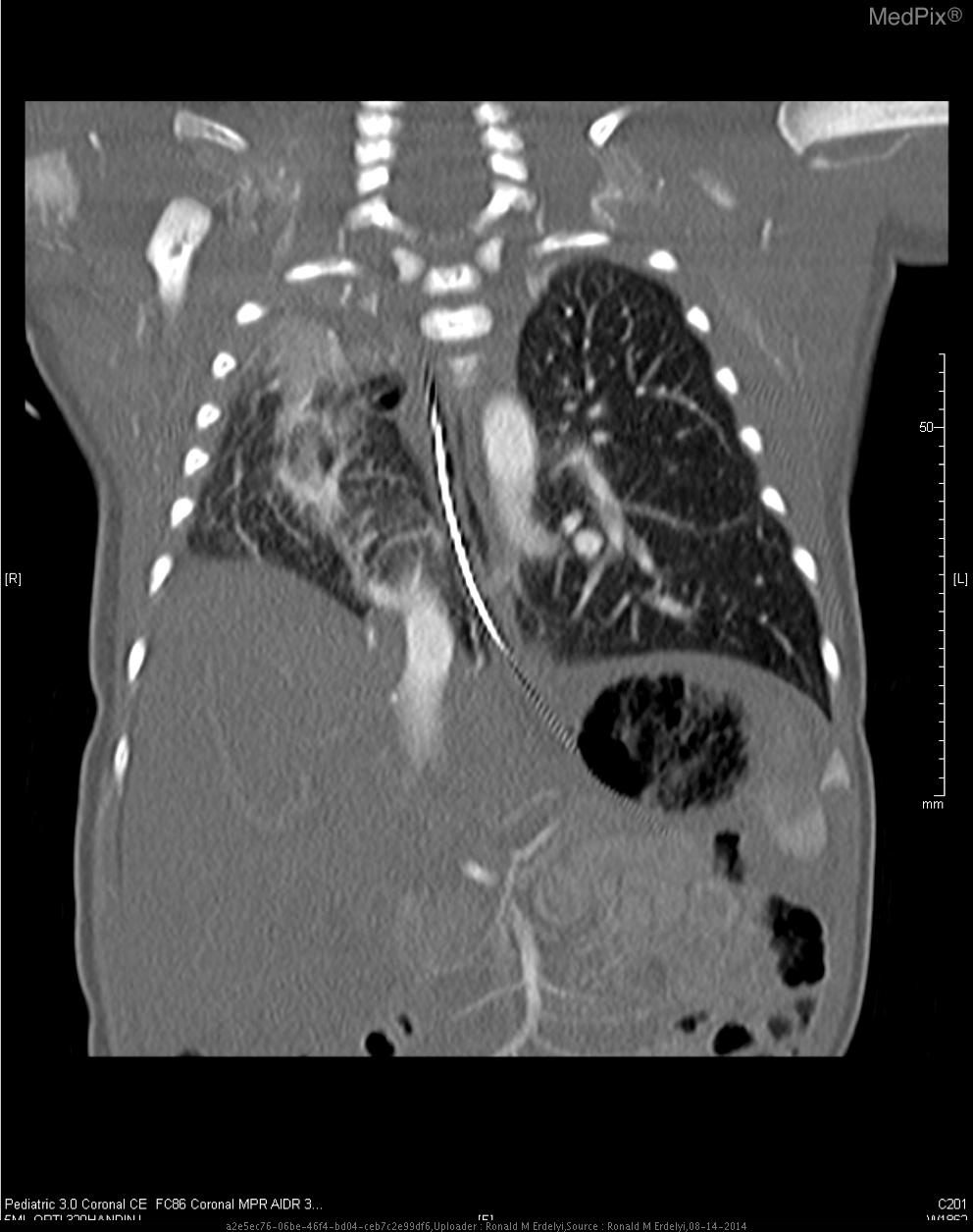
  • Scimitar syndrome
    1. CT with IV contrast shows a hypoplastic right lung with arterial blood supply coming off of the descending aorta, as well as venous drainage directly into inferior vena cava.
https://medpix.nlm.nih.gov/topic?id=6d4e9590-ea5b-4938-be75-e5af30312203
https://medpix.nlm.nih.gov/topic?id=dff03aae-ea33-45c3-8c9a-cf64979526b8

References

Template:WH Template:WS