Donovanosis physical examination: Difference between revisions
Kiran Singh (talk | contribs) No edit summary |
Becca Cohen (talk | contribs) No edit summary |
||
| Line 21: | Line 21: | ||
</gallery> | </gallery> | ||
==Gallery== | |||
<gallery> | |||
Image: Granuloma inguinale11.jpeg| This patient presented with a case of systemically disseminated Donovanosis of the ankle due to C. granulomatis bacteria. <SMALL><SMALL>''[http://phil.cdc.gov/phil/home.asp From Public Health Image Library (PHIL).] ''<ref name=PHIL> {{Cite web | title = Public Health Image Library (PHIL) | url = http://phil.cdc.gov/phil/home.asp}}</ref></SMALL></SMALL> | |||
Image: Granuloma inguinale10.jpeg| This male presented with a penile lesion that was found to be granuloma inguinale, also called “genital ulcerative disease”. <SMALL><SMALL>''[http://phil.cdc.gov/phil/home.asp From Public Health Image Library (PHIL).] ''<ref name=PHIL> {{Cite web | title = Public Health Image Library (PHIL) | url = http://phil.cdc.gov/phil/home.asp}}</ref></SMALL></SMALL> | |||
Image: Granuloma inguinale09.jpeg| This male presented with a penile lesion of roughly 40 days duration that was determined to be granuloma inguinale. <SMALL><SMALL>''[http://phil.cdc.gov/phil/home.asp From Public Health Image Library (PHIL).] ''<ref name=PHIL> {{Cite web | title = Public Health Image Library (PHIL) | url = http://phil.cdc.gov/phil/home.asp}}</ref></SMALL></SMALL> | |||
Image: Granuloma inguinale08.jpeg| This 19 year old woman presented with an perianal granuloma inguinale lesion of about 8 months duration. <SMALL><SMALL>''[http://phil.cdc.gov/phil/home.asp From Public Health Image Library (PHIL).] ''<ref name=PHIL> {{Cite web | title = Public Health Image Library (PHIL) | url = http://phil.cdc.gov/phil/home.asp}}</ref></SMALL></SMALL> | |||
Image: Granuloma inguinale07.jpeg| This patient presented with an ulcerated glans penis due to Donovanosis, or granuloma inguinale. <SMALL><SMALL>''[http://phil.cdc.gov/phil/home.asp From Public Health Image Library (PHIL).] ''<ref name=PHIL> {{Cite web | title = Public Health Image Library (PHIL) | url = http://phil.cdc.gov/phil/home.asp}}</ref></SMALL></SMALL> | |||
Image: Granuloma inguinale06.jpeg| This image depicts an intravaginal view revealing a cervical lesion, which had been diagnosed as a case of Donovanosis, also known as granuloma inguinale. <SMALL><SMALL>''[http://phil.cdc.gov/phil/home.asp From Public Health Image Library (PHIL).] ''<ref name=PHIL> {{Cite web | title = Public Health Image Library (PHIL) | url = http://phil.cdc.gov/phil/home.asp}}</ref></SMALL></SMALL> | |||
Image: Granuloma inguinale05.jpeg| This image depicts the penis of a male with its foreskin retracted, revealing a suppurative lesion involving the glans and prepuce. <SMALL><SMALL>''[http://phil.cdc.gov/phil/home.asp From Public Health Image Library (PHIL).] ''<ref name=PHIL> {{Cite web | title = Public Health Image Library (PHIL) | url = http://phil.cdc.gov/phil/home.asp}}</ref></SMALL></SMALL> | |||
Image: Granuloma inguinale03.jpeg| This image depicts the penis of a male patient who had presented with a lesion located on the lateral preputial skin just proximal to the corona of the glans. The lesion was characterized as a penile granulomata, due to a case of Donovanosis, or granuloma inguinale. <SMALL><SMALL>''[http://phil.cdc.gov/phil/home.asp From Public Health Image Library (PHIL).] ''<ref name=PHIL> {{Cite web | title = Public Health Image Library (PHIL) | url = http://phil.cdc.gov/phil/home.asp}}</ref></SMALL></SMALL> | |||
Image: Granuloma inguinale02.jpeg| This patient showed manifestations of granuloma inguinale, also known as Donovanosis, involving swelling and subcutaneous granulomas of the inguinal lymph nodes, bilaterally. <SMALL><SMALL>''[http://phil.cdc.gov/phil/home.asp From Public Health Image Library (PHIL).] ''<ref name=PHIL> {{Cite web | title = Public Health Image Library (PHIL) | url = http://phil.cdc.gov/phil/home.asp}}</ref></SMALL></SMALL> | |||
Image: Granuloma inguinale01.jpeg| This was a very large erosive cutaneous lesion in the perineal region of this patient, which had been diagnosed as Donovanosis, otherwise known as granuloma inguinale. <SMALL><SMALL>''[http://phil.cdc.gov/phil/home.asp From Public Health Image Library (PHIL).] ''<ref name=PHIL> {{Cite web | title = Public Health Image Library (PHIL) | url = http://phil.cdc.gov/phil/home.asp}}</ref></SMALL></SMALL> | |||
</gallery> | |||
==References== | ==References== | ||
<references/> | <references/> | ||
Revision as of 20:25, 9 June 2015
|
Donovanosis Microchapters |
|
Diagnosis |
|---|
|
Treatment |
|
Case Studies |
|
Donovanosis physical examination On the Web |
|
American Roentgen Ray Society Images of Donovanosis physical examination |
|
Risk calculators and risk factors for Donovanosis physical examination |
Please help WikiDoc by adding more content here. It's easy! Click here to learn about editing.
Editor-In-Chief: C. Michael Gibson, M.S., M.D. [1] Associate Editor(s)-in-Chief: Kalsang Dolma, M.B.B.S.[2] Kiran Singh, M.D. [3]
Overview
Clinically, the disease is commonly characterized as painless, progressive ulcerative lesions without regional lymphadenopathy.
Physical Examination
Clinically, the disease is commonly characterized as painless, progressive ulcerative lesions without regional lymphadenopathy. The lesions are highly vascular (i.e., beefy red appearance) and bleed easily on contact. However, the clinical presentation also can include hypertrophic, necrotic, or sclerotic variants.
-
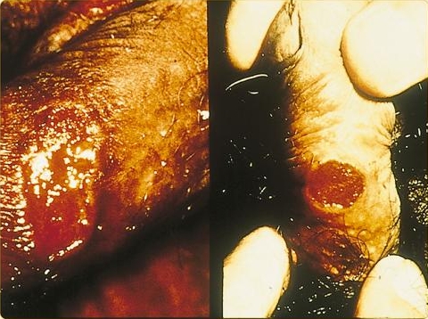
Genital ulcer in a male patient with Donovanosis.
-
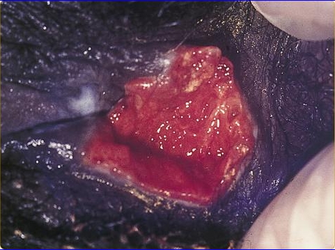
Genital ulcer in a female patient with Donovanosis.


Skin
Genitalia
-
![Donovanosis. Adapted from Dermatology Atlas.[1]](/images/3/38/Donovanosis01.jpg)
Donovanosis. Adapted from Dermatology Atlas.[1]
![Donovanosis. Adapted from Dermatology Atlas.[1]](/index.php/File:Donovanosis01.jpg)
Gallery
-
![This patient presented with a case of systemically disseminated Donovanosis of the ankle due to C. granulomatis bacteria. From Public Health Image Library (PHIL). [2]](/images/4/4a/Granuloma_inguinale11.jpeg)
This patient presented with a case of systemically disseminated Donovanosis of the ankle due to C. granulomatis bacteria. From Public Health Image Library (PHIL). [2]
-
![This male presented with a penile lesion that was found to be granuloma inguinale, also called “genital ulcerative disease”. From Public Health Image Library (PHIL). [2]](/images/3/3c/Granuloma_inguinale10.jpeg)
This male presented with a penile lesion that was found to be granuloma inguinale, also called “genital ulcerative disease”. From Public Health Image Library (PHIL). [2]
-
![This male presented with a penile lesion of roughly 40 days duration that was determined to be granuloma inguinale. From Public Health Image Library (PHIL). [2]](/images/f/f8/Granuloma_inguinale09.jpeg)
This male presented with a penile lesion of roughly 40 days duration that was determined to be granuloma inguinale. From Public Health Image Library (PHIL). [2]
-
![This 19 year old woman presented with an perianal granuloma inguinale lesion of about 8 months duration. From Public Health Image Library (PHIL). [2]](/images/5/50/Granuloma_inguinale08.jpeg)
This 19 year old woman presented with an perianal granuloma inguinale lesion of about 8 months duration. From Public Health Image Library (PHIL). [2]
-
![This patient presented with an ulcerated glans penis due to Donovanosis, or granuloma inguinale. From Public Health Image Library (PHIL). [2]](/images/a/a4/Granuloma_inguinale07.jpeg)
This patient presented with an ulcerated glans penis due to Donovanosis, or granuloma inguinale. From Public Health Image Library (PHIL). [2]
-
![This image depicts an intravaginal view revealing a cervical lesion, which had been diagnosed as a case of Donovanosis, also known as granuloma inguinale. From Public Health Image Library (PHIL). [2]](/images/2/2e/Granuloma_inguinale06.jpeg)
This image depicts an intravaginal view revealing a cervical lesion, which had been diagnosed as a case of Donovanosis, also known as granuloma inguinale. From Public Health Image Library (PHIL). [2]
-
![This image depicts the penis of a male with its foreskin retracted, revealing a suppurative lesion involving the glans and prepuce. From Public Health Image Library (PHIL). [2]](/images/1/10/Granuloma_inguinale05.jpeg)
This image depicts the penis of a male with its foreskin retracted, revealing a suppurative lesion involving the glans and prepuce. From Public Health Image Library (PHIL). [2]
-
![This image depicts the penis of a male patient who had presented with a lesion located on the lateral preputial skin just proximal to the corona of the glans. The lesion was characterized as a penile granulomata, due to a case of Donovanosis, or granuloma inguinale. From Public Health Image Library (PHIL). [2]](/images/2/2a/Granuloma_inguinale03.jpeg)
This image depicts the penis of a male patient who had presented with a lesion located on the lateral preputial skin just proximal to the corona of the glans. The lesion was characterized as a penile granulomata, due to a case of Donovanosis, or granuloma inguinale. From Public Health Image Library (PHIL). [2]
-
![This patient showed manifestations of granuloma inguinale, also known as Donovanosis, involving swelling and subcutaneous granulomas of the inguinal lymph nodes, bilaterally. From Public Health Image Library (PHIL). [2]](/images/8/82/Granuloma_inguinale02.jpeg)
This patient showed manifestations of granuloma inguinale, also known as Donovanosis, involving swelling and subcutaneous granulomas of the inguinal lymph nodes, bilaterally. From Public Health Image Library (PHIL). [2]
-
![This was a very large erosive cutaneous lesion in the perineal region of this patient, which had been diagnosed as Donovanosis, otherwise known as granuloma inguinale. From Public Health Image Library (PHIL). [2]](/images/6/6d/Granuloma_inguinale01.jpeg)
This was a very large erosive cutaneous lesion in the perineal region of this patient, which had been diagnosed as Donovanosis, otherwise known as granuloma inguinale. From Public Health Image Library (PHIL). [2]
![This patient presented with a case of systemically disseminated Donovanosis of the ankle due to C. granulomatis bacteria. From Public Health Image Library (PHIL). [2]](/index.php/File:Granuloma_inguinale11.jpeg)
![This male presented with a penile lesion that was found to be granuloma inguinale, also called “genital ulcerative disease”. From Public Health Image Library (PHIL). [2]](/index.php/File:Granuloma_inguinale10.jpeg)
![This male presented with a penile lesion of roughly 40 days duration that was determined to be granuloma inguinale. From Public Health Image Library (PHIL). [2]](/index.php/File:Granuloma_inguinale09.jpeg)
![This 19 year old woman presented with an perianal granuloma inguinale lesion of about 8 months duration. From Public Health Image Library (PHIL). [2]](/index.php/File:Granuloma_inguinale08.jpeg)
![This patient presented with an ulcerated glans penis due to Donovanosis, or granuloma inguinale. From Public Health Image Library (PHIL). [2]](/index.php/File:Granuloma_inguinale07.jpeg)
![This image depicts an intravaginal view revealing a cervical lesion, which had been diagnosed as a case of Donovanosis, also known as granuloma inguinale. From Public Health Image Library (PHIL). [2]](/index.php/File:Granuloma_inguinale06.jpeg)
![This image depicts the penis of a male with its foreskin retracted, revealing a suppurative lesion involving the glans and prepuce. From Public Health Image Library (PHIL). [2]](/index.php/File:Granuloma_inguinale05.jpeg)
![This image depicts the penis of a male patient who had presented with a lesion located on the lateral preputial skin just proximal to the corona of the glans. The lesion was characterized as a penile granulomata, due to a case of Donovanosis, or granuloma inguinale. From Public Health Image Library (PHIL). [2]](/index.php/File:Granuloma_inguinale03.jpeg)
![This patient showed manifestations of granuloma inguinale, also known as Donovanosis, involving swelling and subcutaneous granulomas of the inguinal lymph nodes, bilaterally. From Public Health Image Library (PHIL). [2]](/index.php/File:Granuloma_inguinale02.jpeg)
![This was a very large erosive cutaneous lesion in the perineal region of this patient, which had been diagnosed as Donovanosis, otherwise known as granuloma inguinale. From Public Health Image Library (PHIL). [2]](/index.php/File:Granuloma_inguinale01.jpeg)