Desmoid tumor CT
|
Desmoid tumor Microchapters |
|
Diagnosis |
|---|
|
Treatment |
|
Case Studies |
Editor-In-Chief: C. Michael Gibson, M.S., M.D. [1] Associate Editor(s)-in-Chief: Faizan Sheraz, M.D. [2]
Overview
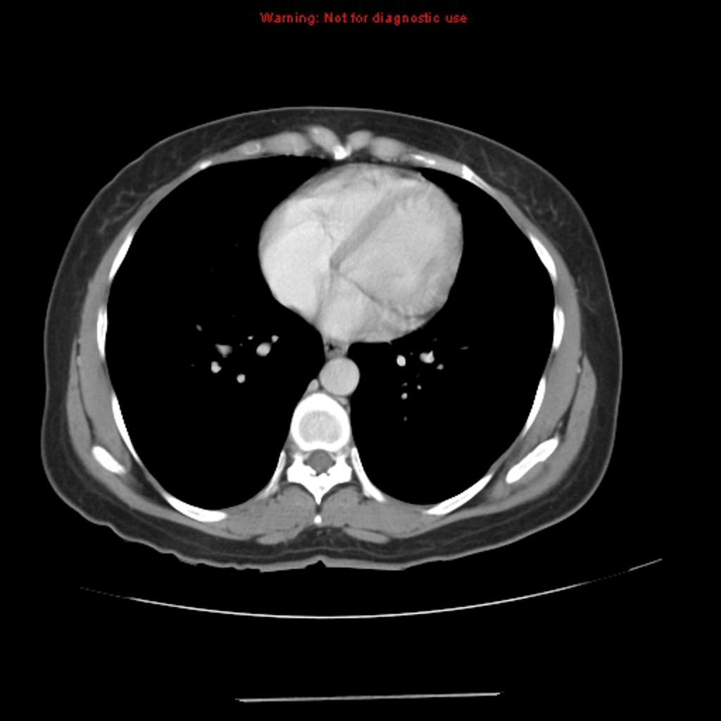
CT scan may be diagnostic for desmoid tumor. On CT scan, desmoid tumor is characterized by a well circumscribed mass that is homogeneously or focally hyperattenuating. Desmoid tumor may demonstrate enhancement following administration of intravenous contrast.[1][2]
CT
CT scan may be diagnostic for desmoid tumor. On CT scan, desmoid tumor is characterized by:
- Well circumscribed mass, although in some cases it may appear more aggressive with ill-defined margins.
- Homogeneously or focally hyperattenuating when compared to soft tissue on the non-contrast scan.
- Most will demonstrate enhancement following administration of intravenous contrast.[1][2]
-
Desmoid tumor of rectus abdominis muscle

Reference
- ↑ 1.0 1.1 Desmoid tumor. Radiopedia(2015) http://radiopaedia.org/articles/aggressive-fibromatosis. Accessed on January 20, 2015
- ↑ 2.0 2.1 Economou, Athanasios; Pitta, Xanthi; Andreadis, Efstathios; Papapavlou, Leonidas; Chrissidis, Thomas (2011). "Desmoid tumor of the abdominal wall: a case report". Journal of Medical Case Reports. 5 (1): 326. doi:10.1186/1752-1947-5-326. ISSN 1752-1947.