Desmoid tumor CT: Difference between revisions
Jump to navigation
Jump to search
No edit summary |
No edit summary |
||
| Line 7: | Line 7: | ||
most desmoid tumors are well circumscribed masses, although in some cases they may appear more aggressive with ill-defined margins | most desmoid tumors are well circumscribed masses, although in some cases they may appear more aggressive with ill-defined margins | ||
most are relatively homogeneously or focally hyperattenuating when compared to soft tissue on the non-contrast scan. | most are relatively homogeneously or focally hyperattenuating when compared to soft tissue on the non-contrast scan. | ||
most will demonstrate enhancement following administration of intravenous contrast. | most will demonstrate enhancement following administration of intravenous contrast.<ref name="radio"> Desmoid tumor. Radiopedia(2015) http://radiopaedia.org/articles/aggressive-fibromatosis. Accessed on January 20, 2015</ref> | ||
<gallery>Rectus-abdominis-muscle-desmoid-tumour.jpg | Desmoid tumor of rectus abdominis muscle</gallery> | <gallery>Rectus-abdominis-muscle-desmoid-tumour.jpg | Desmoid tumor of rectus abdominis muscle</gallery> | ||
Revision as of 15:02, 20 January 2016
|
Desmoid tumor Microchapters |
|
Diagnosis |
|---|
|
Treatment |
|
Case Studies |
Editor-In-Chief: C. Michael Gibson, M.S., M.D. [1] Associate Editor(s)-in-Chief: Faizan Sheraz, M.D. [2]
Overview
CT
most desmoid tumors are well circumscribed masses, although in some cases they may appear more aggressive with ill-defined margins most are relatively homogeneously or focally hyperattenuating when compared to soft tissue on the non-contrast scan. most will demonstrate enhancement following administration of intravenous contrast.[1]
-
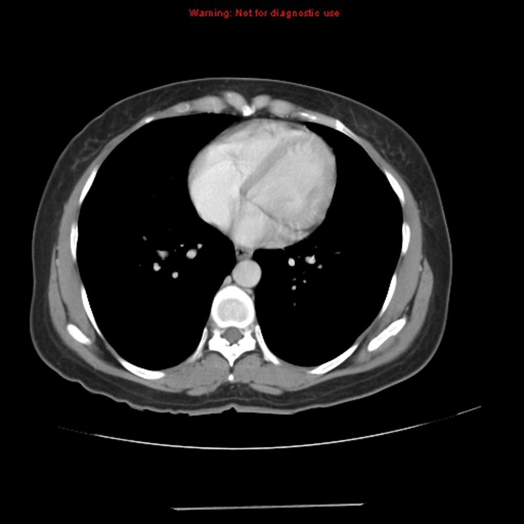
Desmoid tumor of rectus abdominis muscle

Reference
- ↑ Desmoid tumor. Radiopedia(2015) http://radiopaedia.org/articles/aggressive-fibromatosis. Accessed on January 20, 2015