Cirrhosis CT
|
Cirrhosis Microchapters |
|
Diagnosis |
|---|
|
Treatment |
|
Case studies |
|
Cirrhosis CT On the Web |
|
American Roentgen Ray Society Images of Cirrhosis CT |
Editor-In-Chief: C. Michael Gibson, M.S., M.D. [1] ; Associate Editor(s)-in-Chief: Aditya Govindavarjhulla, M.B.B.S. [2]
Overview
Although CT scans are not routinely used in evaluation and diagnosis of cirrhosis, it can show the presence of lobar atrophic and hypertrophic changes in the liver, as well as ascites and varices in advanced disease. CT can also visualize the presence of tumors and blocked bile ducts, as well as evaluate the size of the liver.
CT
Computed tomography is not routinely used in the diagnosis and evaluation of cirrhosis. It is poor at detecting morphologic changes associated with early cirrhosis, but it can accurately demonstrate nodularity and lobar atrophic and hypertrophic changes, as well as ascites and varices in advanced disease. It provides similar information to ultrasonography, but at the expense of radiation and contrast exposure. CT findings may suggest the presence of cirrhosis, but they are not diagnostic. CT portal phase imaging can be used to assess portal vein patency, although flow volume and direction cannot be determined accurately.[1] A computed tomography scan of the abdomen (including the liver, gallbladder, and spleen) can check for tumors and blocked bile ducts and can be used to evaluate liver size and blood flow through the liver.
CT Images
-
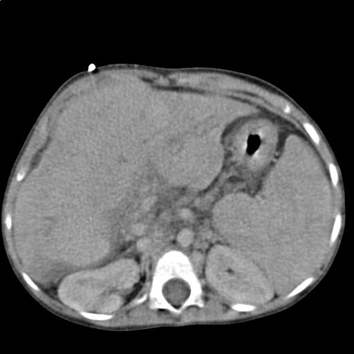
Liver cirrhosis as seen on an axial CT of the abdomen.
