Boerhaave syndrome chest x ray: Difference between revisions
No edit summary |
|||
| (20 intermediate revisions by 4 users not shown) | |||
| Line 1: | Line 1: | ||
__NOTOC__ | __NOTOC__ | ||
{{Boerhaave syndrome}} | {{Boerhaave syndrome}} | ||
{{CMG}} {{AE}} {{DM}} {{SHH}} | {{CMG}} {{AE}} {{DM}}, {{SHH}}, {{Ajay}}, {{FT}} | ||
==Overview== | ==Overview== | ||
An upright anterior-posterior view of the chest is the most useful in early diagnosis. In most patients with [[Boerhaave syndrome]] (BHS) [[chest x-ray]] shows one-sided effusion, [[pneumothorax]], [[hydropneumothorax]], [[pneumomediastinum]] and [[subcutaneous emphysema]]. | |||
==Chest X Ray== | ==Chest X Ray== | ||
* An upright anterior-posterior view of the chest is the most useful in early diagnosis, as most of the patients will reveal an abnormal chest finding after the [[perforation]]. | |||
* The '''Naclerio V-sign''' may be seen on chest [[Radiography|radiograph]] as radiolucent streaks of air seen in the retro-[[cardiac]] region in the V shape.<ref name="pmid28050085">{{cite journal |vauthors=Maurya VK, Sharma P, Ravikumar R, Bhatia M |title=Boerhaave's syndrome |journal=Med J Armed Forces India |volume=72 |issue=Suppl 1 |pages=S105–S107 |year=2016 |pmid=28050085 |pmc=5192176 |doi=10.1016/j.mjafi.2015.12.004 |url=}}</ref> | |||
The usual although unspecific radiographic features of BHS include:<ref name="pmid23493470">{{cite journal |vauthors=Tonolini M, Bianco R |title=Spontaneous esophageal perforation (Boerhaave syndrome): Diagnosis with CT-esophagography |journal=J Emerg Trauma Shock |volume=6 |issue=1 |pages=58–60 |year=2013 |pmid=23493470 |pmc=3589863 |doi=10.4103/0974-2700.106329 |url=}}</ref> | |||
* One-sided [[effusion]] (usually on the left) | |||
* Lung infiltrates | |||
* [[Atelectasis]] | |||
Whereas more specific signs are rarely detected or very subtle:<ref name="pmid28050085">{{cite journal |vauthors=Maurya VK, Sharma P, Ravikumar R, Bhatia M |title=Boerhaave's syndrome |journal=Med J Armed Forces India |volume=72 |issue=Suppl 1 |pages=S105–S107 |year=2016 |pmid=28050085 |pmc=5192176 |doi=10.1016/j.mjafi.2015.12.004 |url=}}</ref><ref name="pmid2730190">{{cite journal |vauthors=Pate JW, Walker WA, Cole FH, Owen EW, Johnson WH |title=Spontaneous rupture of the esophagus: a 30-year experience |journal=Ann. Thorac. Surg. |volume=47 |issue=5 |pages=689–92 |year=1989 |pmid=2730190 |doi= |url=}}</ref><ref name="pmid23493470">{{cite journal |vauthors=Tonolini M, Bianco R |title=Spontaneous esophageal perforation (Boerhaave syndrome): Diagnosis with CT-esophagography |journal=J Emerg Trauma Shock |volume=6 |issue=1 |pages=58–60 |year=2013 |pmid=23493470 |pmc=3589863 |doi=10.4103/0974-2700.106329 |url=}}</ref> | |||
* [[Pneumothorax]] | |||
* [[Hydropneumothorax]] | |||
* [[Pneumomediastinum]] | |||
* [[Widened mediastinum|Mediastinal widening]] | |||
* [[Pneumopericardium]] | |||
* [[Subcutaneous emphysema]] | |||
<gallery> | |||
Image: CXR-BHS.jpg|'''Chest X-ray: Frontal view reveals pneumomediastinum (black arrows). Subcutaneous emphysema (red arrow) along the chest wall, more prominent along the right than left; Source- Radiopaedia''' | |||
Image: CXR BHS-2.jpg|'''Chest X-ray: Boerhaave syndrome- Lateral radiographs subcutaneous emphysema (red arrow) along the chest wall; Source- Radiopaedia''' | |||
</gallery> | |||
==References== | ==References== | ||
Latest revision as of 19:22, 17 February 2021
|
Boerhaave syndrome Microchapters |
|
Diagnosis |
|---|
|
Treatment |
|
Case Studies |
|
Boerhaave syndrome chest x ray On the Web |
|
American Roentgen Ray Society Images of Boerhaave syndrome chest x ray |
|
Risk calculators and risk factors for Boerhaave syndrome chest x ray |
Editor-In-Chief: C. Michael Gibson, M.S., M.D. [1] Associate Editor(s)-in-Chief: Mohamed Diab, MD [2], Shaghayegh Habibi, M.D.[3], Ajay Gade MD[4]], Feham Tariq, MD [5]
Overview
An upright anterior-posterior view of the chest is the most useful in early diagnosis. In most patients with Boerhaave syndrome (BHS) chest x-ray shows one-sided effusion, pneumothorax, hydropneumothorax, pneumomediastinum and subcutaneous emphysema.
Chest X Ray
- An upright anterior-posterior view of the chest is the most useful in early diagnosis, as most of the patients will reveal an abnormal chest finding after the perforation.
- The Naclerio V-sign may be seen on chest radiograph as radiolucent streaks of air seen in the retro-cardiac region in the V shape.[1]
The usual although unspecific radiographic features of BHS include:[2]
- One-sided effusion (usually on the left)
- Lung infiltrates
- Atelectasis
Whereas more specific signs are rarely detected or very subtle:[1][3][2]
- Pneumothorax
- Hydropneumothorax
- Pneumomediastinum
- Mediastinal widening
- Pneumopericardium
- Subcutaneous emphysema
-
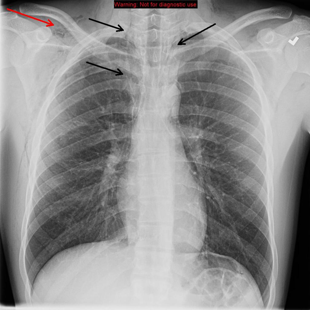
Chest X-ray: Frontal view reveals pneumomediastinum (black arrows). Subcutaneous emphysema (red arrow) along the chest wall, more prominent along the right than left; Source- Radiopaedia
-
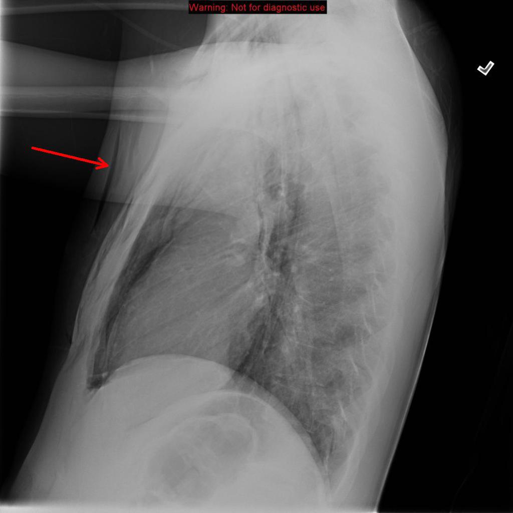
Chest X-ray: Boerhaave syndrome- Lateral radiographs subcutaneous emphysema (red arrow) along the chest wall; Source- Radiopaedia


References
- ↑ 1.0 1.1 Maurya VK, Sharma P, Ravikumar R, Bhatia M (2016). "Boerhaave's syndrome". Med J Armed Forces India. 72 (Suppl 1): S105–S107. doi:10.1016/j.mjafi.2015.12.004. PMC 5192176. PMID 28050085.
- ↑ 2.0 2.1 Tonolini M, Bianco R (2013). "Spontaneous esophageal perforation (Boerhaave syndrome): Diagnosis with CT-esophagography". J Emerg Trauma Shock. 6 (1): 58–60. doi:10.4103/0974-2700.106329. PMC 3589863. PMID 23493470.
- ↑ Pate JW, Walker WA, Cole FH, Owen EW, Johnson WH (1989). "Spontaneous rupture of the esophagus: a 30-year experience". Ann. Thorac. Surg. 47 (5): 689–92. PMID 2730190.