Biliary cystadenoma and cystadenocarcinoma: Difference between revisions
No edit summary |
No edit summary |
||
| Line 36: | Line 36: | ||
==Diagnosis== | ==Diagnosis== | ||
===History and Symptoms=== | |||
The clinical presentation is variable, depending on the size and location of the cyst. Abdominal pain, obstructive jaundice, palpable mass, increasing abdominal girth, nausea, and vomiting are common signs and symptoms. | The clinical presentation is variable, depending on the size and location of the cyst. Abdominal pain, obstructive jaundice, palpable mass, increasing abdominal girth, nausea, and vomiting are common signs and symptoms. | ||
Biliary cystadenomas range in size from 3 to 40 cm. Large cystadenomas may demonstrate mass effect on adjacent organs or may be associated with [[hepatomegaly]]. | Biliary cystadenomas range in size from 3 to 40 cm. Large cystadenomas may demonstrate mass effect on adjacent organs or may be associated with [[hepatomegaly]]. | ||
===Computed Tomography=== | |||
There are no specific imaging features that permit reliable differentiation of biliary cystadenoma from cystadenocarcinoma. | |||
*The CT attenuation of the fluid component in a biliary cystadenoma varies depending on the fluid content. | *The CT attenuation of the fluid component in a biliary cystadenoma varies depending on the fluid content. | ||
| Line 58: | Line 60: | ||
===MRI=== | ===MRI=== | ||
There are no specific imaging features that permit reliable differentiation of biliary cystadenoma from cystadenocarcinoma. | |||
*The MR signal intensity of biliary cystadenoma is variable on both T1- and T2-weighted images, depending on the content of the cyst fluid. | *The MR signal intensity of biliary cystadenoma is variable on both T1- and T2-weighted images, depending on the content of the cyst fluid. | ||
===Ultrasound=== | ===Ultrasound=== | ||
There are no specific imaging features that permit reliable differentiation of biliary cystadenoma from cystadenocarcinoma. | |||
*At US, a biliary cystadenoma appears as a unilocular or multilocular cyst with enhanced through transmission. | *At US, a biliary cystadenoma appears as a unilocular or multilocular cyst with enhanced through transmission. | ||
*Acoustic shadowing may be present from septal or wall calcification. | *Acoustic shadowing may be present from septal or wall calcification. | ||
Revision as of 17:07, 12 September 2012
| Biliary cystadenoma and cystadenocarcinoma | |
 | |
|---|---|
| Biliary cystadenoma (Image courtesy of RadsWiki) |
Editor-In-Chief: C. Michael Gibson, M.S., M.D. [1]
Overview
Biliary cystadenomas are uncommon unilocular or multilocular cystic neoplasms that may occur within the liver (infrequently found in the extrahepatic biliary tree and gallbladder).
Although biliary cystadenomas are benign tumors, they may recur after excision and have potential to develop into biliary cystadenocarcinoma.
Pathophysiology
Microscopic Pathology
At histologic analysis, cystadenomas have multiple loculations lined by cuboidal or columnar epithelium that resembles biliary epithelium.
Differenting Biliary cystadenoma and cystadenocarcinoma from other Diseases
The differential diagnosis principally includes
Epidemiology and Demographics
Cystadenomas occur predominantly in middle-aged women.
Diagnosis
History and Symptoms
The clinical presentation is variable, depending on the size and location of the cyst. Abdominal pain, obstructive jaundice, palpable mass, increasing abdominal girth, nausea, and vomiting are common signs and symptoms.
Biliary cystadenomas range in size from 3 to 40 cm. Large cystadenomas may demonstrate mass effect on adjacent organs or may be associated with hepatomegaly.
Computed Tomography
There are no specific imaging features that permit reliable differentiation of biliary cystadenoma from cystadenocarcinoma.
- The CT attenuation of the fluid component in a biliary cystadenoma varies depending on the fluid content.
- Higher attenuation may indicate recent hemorrhage.
- Calcifications that may be present in the septa or cyst wall are typically more apparent with CT than other imaging modalities.
- Septa may enhance with contrast material.
(Images courtesy of RadsWiki)
-
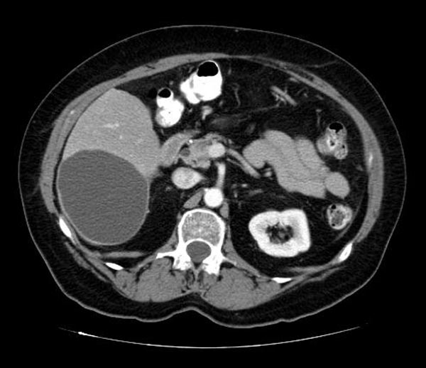
CT image demonstates a biliary cystadenoma
-
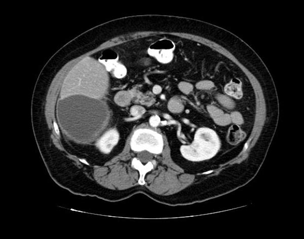
CT image demonstates a biliary cystadenoma

MRI
There are no specific imaging features that permit reliable differentiation of biliary cystadenoma from cystadenocarcinoma.
- The MR signal intensity of biliary cystadenoma is variable on both T1- and T2-weighted images, depending on the content of the cyst fluid.
Ultrasound
There are no specific imaging features that permit reliable differentiation of biliary cystadenoma from cystadenocarcinoma.
- At US, a biliary cystadenoma appears as a unilocular or multilocular cyst with enhanced through transmission.
- Acoustic shadowing may be present from septal or wall calcification.
- The cyst fluid may contain low-level echoes from blood products, mucin, or proteinaceous fluid. Serous and bilious cyst fluid is generally anechoic.
- Echogenic mural nodules and papillary projections may be present.
Related Chapters
- Malignant hepatic neoplasms
- Benign hepatic neoplasms
External Links
Reference
- Angela D. Levy, Linda A. Murakata, Robert M. Abbott, and Charles A. Rohrmann, Jr. From the Archives of the AFIP: Benign Tumors and Tumorlike Lesions of the Gallbladder and Extrahepatic Bile Ducts: Radiologic-Pathologic Correlation. RadioGraphics 2002 22: 387-413.