Ascariasis pathophysiology: Difference between revisions
No edit summary |
No edit summary |
||
| Line 3: | Line 3: | ||
==Life cycle== | ==Life cycle== | ||
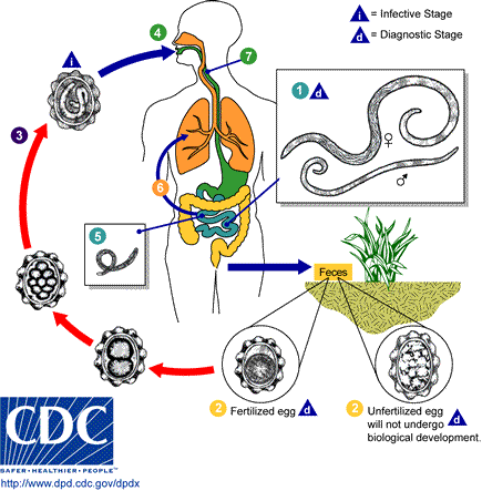
[[Image:Ascariasis LifeCycle - CDC Division of Parasitic Diseases.gif|thumb|left|300px|Adult worms (1) live in the lumen of the small intestine. A female may produce approximately 200,000 eggs per day, which are passed with the feces (2). Unfertilized eggs may be ingested but are not infective. Fertile eggs embryonate and become infective after 18 days to several weeks (3), depending on the environmental conditions (optimum: moist, warm, shaded soil). After infective eggs are swallowed (4), the larvae hatch (5), invade the intestinal mucosa, and are carried via the portal, then systemic circulation to the lungs . The larvae mature further in the lungs (6) (10 to 14 days), penetrate the alveolar walls, ascend the bronchial tree to the throat, and are swallowed (7). Upon reaching the small intestine, they develop into adult worms (8). Between 2 and 3 months are required from ingestion of the infective eggs to oviposition by the adult female. Adult worms can live 1 to 2 years.]]First appearance of eggs in stools is 60-70 days. In larval ascariasis, symptoms occur 4-16 days after infection. The final symptoms are gastrointestinal discomfort, colic and vomiting, fever; observation of live worms in stools. Some patients may have pulmonary symptoms or neurological disorders during migration of the larvae. However there are generally few or no symptoms. A bolus of worms may obstruct the intestine; migrating larvae may cause pneumonitis and [[eosinophilia]]. | [[Image:Ascariasis LifeCycle - CDC Division of Parasitic Diseases.gif|thumb|left|300px|Adult worms (1) live in the lumen of the small intestine. A female may produce approximately 200,000 eggs per day, which are passed with the feces (2). Unfertilized eggs may be ingested but are not infective. Fertile eggs embryonate and become infective after 18 days to several weeks (3), depending on the environmental conditions (optimum: moist, warm, shaded soil). After infective eggs are swallowed (4), the larvae hatch (5), invade the intestinal mucosa, and are carried via the portal, then systemic circulation to the lungs . The larvae mature further in the lungs (6) (10 to 14 days), penetrate the alveolar walls, ascend the bronchial tree to the throat, and are swallowed (7). Upon reaching the small intestine, they develop into adult worms (8). Between 2 and 3 months are required from ingestion of the infective eggs to oviposition by the adult female. Adult worms can live 1 to 2 years.]]First appearance of eggs in stools is 60-70 days. In larval ascariasis, symptoms occur 4-16 days after infection. The final symptoms are gastrointestinal discomfort, colic and vomiting, fever; observation of live worms in stools. Some patients may have pulmonary symptoms or neurological disorders during migration of the larvae. However there are generally few or no symptoms. A bolus of worms may obstruct the intestine; migrating larvae may cause pneumonitis and [[eosinophilia]]. | ||
==Source== | |||
The source of transmission is from soil and vegetation on which fecal matter containing eggs has been deposited. Ingestion of infective eggs from soil contaminated with human feces or transmission and contaminated vegetables and water is the primary route of infection. Intimate contact with pets which have been in contact with contaminated soil may result in infection, while pets which are infested themselves by a different type of roundworm can cause infection with that type of worm (Toxocara canis, etc) as occasionally occurs with groomers. | |||
Transmission also comes through municipal recycling of wastewater into crop fields. This is quite common in emerging industrial economies, and poses serious risks for not only local crop sales but also exports of contaminated vegetables. A 1986 outbreak of ascariasis in Italy was traced to irresponsible wastewater recycling used to grow Balkan vegetable exports. | |||
Transmission from human to human by direct contact is impossible.[http://www.cdc.gov/ncidod/dpd/parasites/ascaris/factsht_ascaris.htm#contagious] | |||
==References== | ==References== | ||
Revision as of 21:14, 24 January 2012
|
Ascariasis Microchapters |
|
Diagnosis |
|---|
|
Treatment |
|
Case Studies |
|
Ascariasis pathophysiology On the Web |
|
American Roentgen Ray Society Images of Ascariasis pathophysiology |
|
Risk calculators and risk factors for Ascariasis pathophysiology |
Editor-In-Chief: C. Michael Gibson, M.S., M.D. [1]; Associate Editor-In-Chief: Imtiaz Ahmed Wani, M.B.B.S
Life cycle

First appearance of eggs in stools is 60-70 days. In larval ascariasis, symptoms occur 4-16 days after infection. The final symptoms are gastrointestinal discomfort, colic and vomiting, fever; observation of live worms in stools. Some patients may have pulmonary symptoms or neurological disorders during migration of the larvae. However there are generally few or no symptoms. A bolus of worms may obstruct the intestine; migrating larvae may cause pneumonitis and eosinophilia.
Source
The source of transmission is from soil and vegetation on which fecal matter containing eggs has been deposited. Ingestion of infective eggs from soil contaminated with human feces or transmission and contaminated vegetables and water is the primary route of infection. Intimate contact with pets which have been in contact with contaminated soil may result in infection, while pets which are infested themselves by a different type of roundworm can cause infection with that type of worm (Toxocara canis, etc) as occasionally occurs with groomers.
Transmission also comes through municipal recycling of wastewater into crop fields. This is quite common in emerging industrial economies, and poses serious risks for not only local crop sales but also exports of contaminated vegetables. A 1986 outbreak of ascariasis in Italy was traced to irresponsible wastewater recycling used to grow Balkan vegetable exports.
Transmission from human to human by direct contact is impossible.[2]
References
de:Spulwurm hu:Orsóférgek io:Askaridiko id:Askariasis it:Ascaridiasi nl:Spoelworm ps:اسکاريس لومبريکويډېس sk:Hlísta detská