|
|
| Line 4: |
Line 4: |
|
| |
|
| ==Overview== | | ==Overview== |
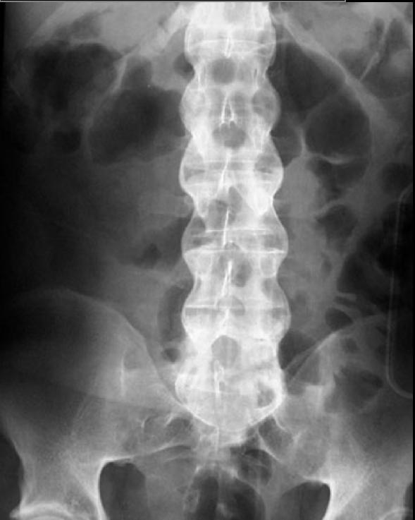
| A clinical examination and [[x-ray]]s of the spine, which show characteristic spinal changes such as [[Sacroiliac joint|sacroiliitis]], are the major diagnostic tools for ankylosing spondylitis. The findings on an x-ray that are consistent with a diagnosis of ankylosing spondilitis are the presence of subchondral erosions, [[sclerosis]], proliferation on the iliac side of SI joints, and squaring of the [[vertebral body]].
| |
|
| |
|
| ==X Ray== | | ==X Ray== |
| [[Image:Morbus Bechterew.jpg|left|thumb|200px|Ankylosing spondylitis (Morbus Bechterew)]] | | [[Image:Morbus Bechterew.jpg|left|thumb|200px|Ankylosing spondylitis (Morbus Bechterew)]] |
| * Indistinct joints | | * |
| * Joints widen before narrow
| |
| * Subchondral erosions, sclerosis, and proliferation on iliac side of SI joints
| |
| * At endstage, sacroiliac joint may be a thin line or not visible
| |
| * In the spine, early spondylitis is characterized by small erosions at the corners of vertebral bodies with reactive [[sclerosis]]
| |
| * Squaring of the vertebral body
| |
| * [[Syndesmophyte]] formation, with bridging of the corners of one vertebra to another
| |
| * [[Ossification]] of paravertebral connective tissue fibers, including posterior interspinous ligaments as well as linking of spinous processes leads to an appearance of a solid midline vertical dense line on AP projection
| |
| * May see associated [[pseudoarthroses]] (discovertebral destruction with adjacent sclerosis) and enthesopathic changes (ill-defined erosions with adjacent sclerosis at sites of ligamentous and tendenous attachments)
| |
| * Hip involvement is generally bilateral and symmetric, with uniform joint space narrowing, axial migration of the femoral head, and a collar of [[osteophyte]]s at the femoral head-neck junction
| |
| * Knees demonstrate uniform joint space narrowing with bony proliferation
| |
| * Hands are generally involved asymmetrically, with smaller, shallower erosions and marginal [[periostitis]].
| |
| * Radiographs of the lungs may demonstrate progressive fibrosis and [[bullous]] changes at the apices. These lesions may resemble TB infection and bullae may become infected.
| |
| <gallery> | | <gallery> |
| Image:Ankylosing-spondylitis-001.jpg|Bamboo Spine | | Image:Ankylosing-spondylitis-001.jpg|Bamboo Spine |
| </gallery> | | </gallery> |
|
| |
| A drawback of X-ray diagnosis is that signs and symptoms of AS have usually been established as long as 8-10 years prior to X-ray evident changes occurring on a plain film X-ray, which means a delay of as long as 10 years before adequate therapies can be introduced.
| |
|
| |
|
| ==References== | | ==References== |