Androgen insensitivity syndrome pathophysiology: Difference between revisions
| Line 44: | Line 44: | ||
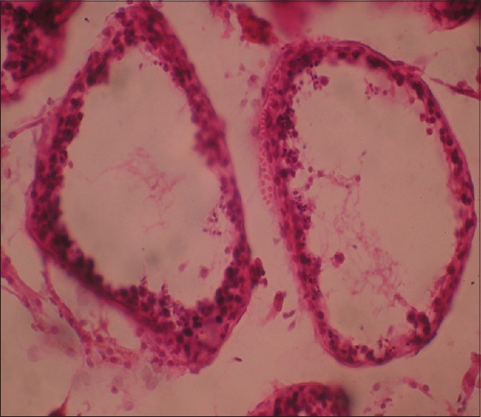
*Histopathology shows two testes with atrophic [[seminiferous tubules]] containing only [[sertoli cells]], associated to a [[leydig cells]] [[hyperplasia]].<ref name="pmid26301004">{{cite journal| author=Lachiri B, Hakimi I, Boudhas A, Guelzim K, Kouach J, Oukabli M et al.| title=[Complete androgen insensitivity syndrome: report of two cases and review of literature]. | journal=Pan Afr Med J | year= 2015 | volume= 20 | issue= | pages= 400 | pmid=26301004 | doi=10.11604/pamj.2015.20.400.6760 | pmc=4524922 | url=https://www.ncbi.nlm.nih.gov/entrez/eutils/elink.fcgi?dbfrom=pubmed&tool=sumsearch.org/cite&retmode=ref&cmd=prlinks&id=26301004 }} </ref> | *Histopathology shows two testes with atrophic [[seminiferous tubules]] containing only [[sertoli cells]], associated to a [[leydig cells]] [[hyperplasia]].<ref name="pmid26301004">{{cite journal| author=Lachiri B, Hakimi I, Boudhas A, Guelzim K, Kouach J, Oukabli M et al.| title=[Complete androgen insensitivity syndrome: report of two cases and review of literature]. | journal=Pan Afr Med J | year= 2015 | volume= 20 | issue= | pages= 400 | pmid=26301004 | doi=10.11604/pamj.2015.20.400.6760 | pmc=4524922 | url=https://www.ncbi.nlm.nih.gov/entrez/eutils/elink.fcgi?dbfrom=pubmed&tool=sumsearch.org/cite&retmode=ref&cmd=prlinks&id=26301004 }} </ref> | ||
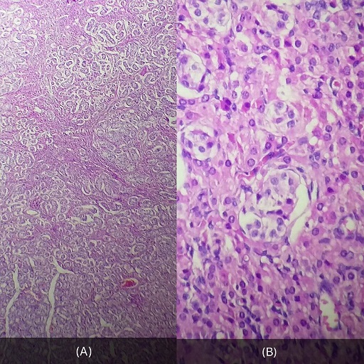
*On histological examination, the well-limited nodule circumscribed by a thin capsule consists of atrophic servolian tubes with a very small interstitial tissue with rare [[leydig cells]]. This nodule corresponds to a well differentiated tumor with [[sertoli-leydig cells]]. <ref name="pmid28270903">{{cite journal| author=Souhail R, Amine S, Nadia A, Tarik K, Khalid EK, Abdellatif K et al.| title=Complete androgen insensitivity syndrome or testicular feminization: review of literature based on a case report. | journal=Pan Afr Med J | year= 2016 | volume= 25 | issue= | pages= 199 | pmid=28270903 | doi=10.11604/pamj.2016.25.199.10758 | pmc=5326263 | url=https://www.ncbi.nlm.nih.gov/entrez/eutils/elink.fcgi?dbfrom=pubmed&tool=sumsearch.org/cite&retmode=ref&cmd=prlinks&id=28270903 }} </ref> | *On histological examination, the well-limited nodule circumscribed by a thin capsule consists of atrophic servolian tubes with a very small interstitial tissue with rare [[leydig cells]]. This nodule corresponds to a well differentiated tumor with [[sertoli-leydig cells]]. <ref name="pmid28270903">{{cite journal| author=Souhail R, Amine S, Nadia A, Tarik K, Khalid EK, Abdellatif K et al.| title=Complete androgen insensitivity syndrome or testicular feminization: review of literature based on a case report. | journal=Pan Afr Med J | year= 2016 | volume= 25 | issue= | pages= 199 | pmid=28270903 | doi=10.11604/pamj.2016.25.199.10758 | pmc=5326263 | url=https://www.ncbi.nlm.nih.gov/entrez/eutils/elink.fcgi?dbfrom=pubmed&tool=sumsearch.org/cite&retmode=ref&cmd=prlinks&id=28270903 }} </ref> | ||
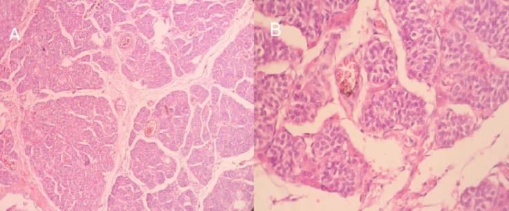
*The gonads histopathological examination show the following: <ref name="pmid25395750">{{cite journal |vauthors=Bhaskararao G, Himabindu Y, Nayak SR, Sriharibabu M |title=Laparoscopic gonedectomy in a case of complete androgen insensitivity syndrome |journal=J Hum Reprod Sci |volume=7 |issue=3 |pages=221–3 |year=2014 |pmid=25395750 |pmc=4229800 |doi=10.4103/0974-1208.142498 |url=}}</ref> | *The gonads on histopathological examination show the following: <ref name="pmid25395750">{{cite journal |vauthors=Bhaskararao G, Himabindu Y, Nayak SR, Sriharibabu M |title=Laparoscopic gonedectomy in a case of complete androgen insensitivity syndrome |journal=J Hum Reprod Sci |volume=7 |issue=3 |pages=221–3 |year=2014 |pmid=25395750 |pmc=4229800 |doi=10.4103/0974-1208.142498 |url=}}</ref> | ||
:*Thickened tunica albuginea | :*Thickened tunica albuginea | ||
:*Seminiferous tubules with primary and secondary spermatogonia and sertoli cells | :*Seminiferous tubules with primary and secondary spermatogonia and sertoli cells | ||
Revision as of 03:05, 24 August 2017
|
Androgen insensitivity syndrome Microchapters |
|
Differentiating Androgen insensitivity syndrome from other Diseases |
|---|
|
Diagnosis |
|
Treatment |
|
Case Studies |
|
Androgen insensitivity syndrome pathophysiology On the Web |
|
American Roentgen Ray Society Images of Androgen insensitivity syndrome pathophysiology |
|
Directions to Hospitals Treating Androgen insensitivity syndrome |
|
Risk calculators and risk factors for Androgen insensitivity syndrome pathophysiology |
Editor-In-Chief: C. Michael Gibson, M.S., M.D. [1]; Associate Editor(s)-in-Chief: Aravind Reddy Kothagadi M.B.B.S[2]
Overview
It is thought that Androgen insensitivity syndrome is caused due to hormone resistance which may be due to defective androgen receptor (AR) function by either abnormal androgen receptor (AR) binding, decreased receptor binding, or impaired androgen receptor (AR) binding. AIS is an X linked disorder. The development of Androgen insensitivity syndrome is a result of genetic mutations of the androgen receptor (AR) gene located on the chromosome Xq11-12. Associated conditions include primary amenorrhea, infertility and dyspareunia.
Pathophysiology
Pathogenesis
- It is thought that Androgen insensitivity syndrome is caused due to hormone resistance which may be due to defective androgen receptor (AR) function by either abnormal androgen receptor (AR) binding, decreased receptor binding, or impaired androgen receptor (AR) binding. [1] [2] [3] [4]
- A spectrum of phenotypes may be caused due to missense mutations in the androgen receptor (AR) protein. The two important domains of the receptor protein such as DBD and LDB domains are the ones wherein the most frequent missense mutations are found. [5] [6]
- The phenotype variability impacts and translates the degree to which ligand-binding and receptor functions are disrupted by different substitutions. [5] [7]
- The genetic background has an influence on the resulting phenotype as a result of the same mutation which may lead to different forms of AIS within a family. [5] [8]
- Mutations in the androgen receptor (AR) gene helps as a trusted tool for the diagnosis and molecular subclassification of AIS. The resulting phenotype is affected by the kind of amino acid substitution occuring due to mutation. [5]
- Information regarding the mutation in the androgen receptor (AR) and its functional consequences helps in determining the genotype-phenotype correlation, to improve and better manage the cases of male pseudohermaphroditism pertaining to surgery of the genitalia, gonadectomy and gender assignment. [5]
Genetics
- AIS is an X linked disorder. [9]
- A high proportion of De novo mutations arise after the zygote stage which occur at a high rate within the androgen receptor (AR) gene. [9]
- Spontaneous mutations in the androgen receptor (AR) gene results in Androgen insensitivity syndrome even without any family history. [9]
- The development of Androgen insensitivity syndrome is a result of genetic mutations of the androgen receptor (AR) gene located on the chromosome Xq11-12. [5] [9]
- Different mutations in the androgen receptor (AR) gene leads to varied clinical phenotypes. [10] [11]
- There have been more than 800 mutations in the androgen receptor (AR) gene reported in AIS patients. [12]
Associated Conditions
Gross Pathology
- Complete androgen insensitivity syndrome in a 30 years old woman presenting with primary amenorrhea.[13]. [14]
-
Front and side view of the patient [13]
-
Normal female morphotype but absence of pubic and axillary hair [14]
-
Clinical aspect of the vagina [13]
-
Intra- abdominal testes - Laparoscopic aspect [13]
-
The excised testis - Macroscopic aspect [13]
Microscopic Pathology
- Histopathology shows two testes with atrophic seminiferous tubules containing only sertoli cells, associated to a leydig cells hyperplasia.[14]
- On histological examination, the well-limited nodule circumscribed by a thin capsule consists of atrophic servolian tubes with a very small interstitial tissue with rare leydig cells. This nodule corresponds to a well differentiated tumor with sertoli-leydig cells. [13]
- The gonads on histopathological examination show the following: [15]
- Thickened tunica albuginea
- Seminiferous tubules with primary and secondary spermatogonia and sertoli cells
- Intertubular leydig cells were seen along with peritubular fibrosis


References
- ↑ Flier, Jeffrey S.; Underhill, Lisa H.; Griffin, James E. (1992). "Androgen Resistance — The Clinical and Molecular Spectrum". New England Journal of Medicine. 326 (9): 611–618. doi:10.1056/NEJM199202273260906. ISSN 0028-4793.
- ↑ Pagon RA, Adam MP, Ardinger HH, Wallace SE, Amemiya A, Bean L, Bird TD, Ledbetter N, Mefford HC, Smith R, Stephens K, Gottlieb B, Trifiro MA. PMID 20301602. Vancouver style error: initials (help); Missing or empty
|title=(help) - ↑ Brown, Terry R.; Maes, Marc; Rothwell, Stephen W.; Migeon, Claude J. (1982). "Human Complete Androgen Insensitivity with Normal Dihydrotestosterone Receptor Binding Capacity in Cultured Genital Skin Fibroblasts: Evidence for a Qualitative Abnormality of the Receptor*". The Journal of Clinical Endocrinology & Metabolism. 55 (1): 61–69. doi:10.1210/jcem-55-1-61. ISSN 0021-972X.
- ↑ Griffin, James E. (1979). "Testicular Feminization Associated with a Thermolabile Androgen Receptor in Cultured Human Fibroblasts". Journal of Clinical Investigation. 64 (6): 1624–1631. doi:10.1172/JCI109624. ISSN 0021-9738.
- ↑ 5.0 5.1 5.2 5.3 5.4 5.5 Kota SK, Gayatri K, Kota SK, Jammula S (2013). "Genetic analysis of a family with complete androgen insensitivity syndrome". Indian J Hum Genet. 19 (3): 355–7. doi:10.4103/0971-6866.120820. PMC 3841565. PMID 24339553.
- ↑ Brinkmann AO, Faber PW, van Rooij HC, Kuiper GG, Ris C, Klaassen P, van der Korput JA, Voorhorst MM, van Laar JH, Mulder E (1989). "The human androgen receptor: domain structure, genomic organization and regulation of expression". J. Steroid Biochem. 34 (1–6): 307–10. PMID 2626022.
- ↑ McPhaul MJ, Marcelli M, Tilley WD, Griffin JE, Wilson JD (1991). "Androgen resistance caused by mutations in the androgen receptor gene". FASEB J. 5 (14): 2910–5. PMID 1752359.
- ↑ Evans BA, Hughes IA, Bevan CL, Patterson MN, Gregory JW (1997). "Phenotypic diversity in siblings with partial androgen insensitivity syndrome". Arch. Dis. Child. 76 (6): 529–31. PMC 1717223. PMID 9245853.
- ↑ 9.0 9.1 9.2 9.3 Akella RR (2017). "Mutational Analysis of Androgen Receptor Gene in Two Families with Androgen Insensitivity". Indian J Endocrinol Metab. 21 (4): 520–523. doi:10.4103/ijem.IJEM_345_16. PMC 5477437. PMID 28670533.
- ↑ Li L, Liu WM, Liu MX, Zheng SQ, Zhang JX, Che FY, Liu SG (2017). "A missense mutation in the androgen receptor gene causing androgen insensitivity syndrome in a Chinese family". Asian J. Androl. 19 (2): 260–261. doi:10.4103/1008-682X.172647. PMC 5312231. PMID 26806084.
- ↑ Brinkmann, Albert O. (2001). "Molecular basis of androgen insensitivity". Molecular and Cellular Endocrinology. 179 (1–2): 105–109. doi:10.1016/S0303-7207(01)00466-X. ISSN 0303-7207.
- ↑ Hughes IA, Werner R, Bunch T, Hiort O (2012). "Androgen insensitivity syndrome". Semin Reprod Med. 30 (5): 432–42. doi:10.1055/s-0032-1324728. PMID 23044881.
- ↑ 13.0 13.1 13.2 13.3 13.4 13.5 13.6 Souhail R, Amine S, Nadia A, Tarik K, Khalid EK, Abdellatif K, Ahmed A (2016). "Complete androgen insensitivity syndrome or testicular feminization: review of literature based on a case report". Pan Afr Med J. 25: 199. doi:10.11604/pamj.2016.25.199.10758. PMC 5326263. PMID 28270903.
- ↑ 14.0 14.1 14.2 14.3 Lachiri B, Hakimi I, Boudhas A, Guelzim K, Kouach J, Oukabli M, Rahali DM, Dehayni M (2015). "[Complete androgen insensitivity syndrome: report of two cases and review of literature]". Pan Afr Med J (in French). 20: 400. doi:10.11604/pamj.2015.20.400.6760. PMC 4524922. PMID 26301004.
- ↑ 15.0 15.1 Bhaskararao G, Himabindu Y, Nayak SR, Sriharibabu M (2014). "Laparoscopic gonedectomy in a case of complete androgen insensitivity syndrome". J Hum Reprod Sci. 7 (3): 221–3. doi:10.4103/0974-1208.142498. PMC 4229800. PMID 25395750.
![Front and side view of the patient [13]](/images/e/e3/AIS_-_Front_and_side_view_of_the_patient.jpg)
![Normal female morphotype but absence of pubic and axillary hair [14]](/images/5/5f/AIS_-_absence_of_pubic_and_axillary_hair.jpg)
![Clinical aspect of the vagina [13]](/images/e/eb/AIS_-_Clinical_aspect_of_the_vagina.jpg)
![Intra- abdominal testes - Laparoscopic aspect [13]](/images/d/d8/AIS_-_Intra-_abdominal_testes_-_laparoscopic_aspect.jpg)
![The excised testis - Macroscopic aspect [13]](/images/7/7d/AIS_-_The_excised_testis_-_Macroscopic_aspect.jpg)