Amyloidosis CT scan: Difference between revisions
Jump to navigation
Jump to search
(Created page with "__NOTOC__ {{Amyloidosis}} {{CMG}} {{shyam}}; {{AE}} {{SHH}}{{Sab}} ==Overview== CT can be done to assess for amyloid deposition in particular organs. It can also be done to r...") |
No edit summary |
||
| (2 intermediate revisions by 2 users not shown) | |||
| Line 4: | Line 4: | ||
==Overview== | ==Overview== | ||
CT can be done to assess for amyloid deposition in particular organs. It can also be done to rule out other causes of organ dysfunction. However, MRI is more sensitive than CT in the diagnosis of amyloidosis. | [[Computed tomography|CT scan]] can be done to assess for amyloid deposition in particular organs. It can also be done to rule out other causes of organ dysfunction. However, MRI is more sensitive than CT in the diagnosis of amyloidosis. | ||
==CT== | ==CT scan== | ||
In hepatic amyloidosis | In hepatic amyloidosis, [[Computed tomography|CT scan]] findings may include: | ||
* Liver enlargement with heterogeneous decreased attenuation | *[[Liver]] enlargement with [[heterogeneous]] decreased attenuation | ||
* Asymmetric and triangular hepatomegaly with the apex at the falciform ligament (due to mild atrophic change of the lateral border of both hepatic lobes) | * Asymmetric and triangular [[hepatomegaly]] with the apex at the [[falciform ligament]] (due to mild [[Atrophy|atrophic]] change of the lateral border of both [[Liver|hepatic]] lobes) | ||
* Parenchyma calcification (rare) | *[[Parenchyma|Parenchymal]] [[calcification]] (rare) | ||
In renal amyloidosis | In [[Kidney|renal]] amyloidosis, [[Computed tomography|CT scan]] findings may include: | ||
* Kidney enlargement with heterogeneous decreased attenuation | *[[Kidney]] enlargement with [[heterogeneous]] decreased attenuation | ||
* Parenchyma calcification (rare) | *[[Parenchyma|Parenchymal]] [[calcification]] (rare) | ||
In cardiac amyloidosis | In [[Heart|cardiac]] amyloidosis, [[Computed tomography|CT scan]] findings may include<ref name="pmid24847009">{{cite journal| author=Falk RH, Quarta CC, Dorbala S| title=How to image cardiac amyloidosis. | journal=Circ Cardiovasc Imaging | year= 2014 | volume= 7 | issue= 3 | pages= 552-62 | pmid=24847009 | doi=10.1161/CIRCIMAGING.113.001396 | pmc=4118308 | url=https://www.ncbi.nlm.nih.gov/entrez/eutils/elink.fcgi?dbfrom=pubmed&tool=sumsearch.org/cite&retmode=ref&cmd=prlinks&id=24847009 }} </ref>: | ||
* Heart enlargement with heterogeneous decreased attenuation | *[[Heart]] enlargement with [[heterogeneous]] decreased attenuation | ||
* Cardiac calcifications | *[[Heart|Cardiac]] [[Calcification|calcifications]] | ||
* Pericardial effusion (rare) | *[[Pericardial effusion]] (rare) | ||
===Images=== | ===Images=== | ||
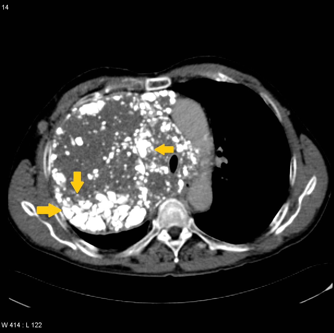
[[File:Amyloidoma-mediastinal-1.jpg|300px|left|thumb| CT image showing mediastinal amyloidosis (yellow arrows). Case courtesy of Dr Natalie Yang, Radiopaedia.org, rID: 6711]] | [[File:Amyloidoma-mediastinal-1.jpg|300px|left|thumb| CT image showing mediastinal amyloidosis (yellow arrows). Case courtesy of Dr Natalie Yang, Radiopaedia.org, rID: 6711]] | ||
Latest revision as of 19:37, 7 November 2019
|
Amyloidosis Microchapters |
|
Diagnosis |
|---|
|
Treatment |
|
Case Studies |
|
Amyloidosis CT scan On the Web |
|
American Roentgen Ray Society Images of Amyloidosis CT scan |
Editor-In-Chief: C. Michael Gibson, M.S., M.D. [1] Shyam Patel [2]; Associate Editor(s)-in-Chief: Shaghayegh Habibi, M.D.[3]Sabawoon Mirwais, M.B.B.S, M.D.[4]
Overview
CT scan can be done to assess for amyloid deposition in particular organs. It can also be done to rule out other causes of organ dysfunction. However, MRI is more sensitive than CT in the diagnosis of amyloidosis.
CT scan
In hepatic amyloidosis, CT scan findings may include:
- Liver enlargement with heterogeneous decreased attenuation
- Asymmetric and triangular hepatomegaly with the apex at the falciform ligament (due to mild atrophic change of the lateral border of both hepatic lobes)
- Parenchymal calcification (rare)
In renal amyloidosis, CT scan findings may include:
- Kidney enlargement with heterogeneous decreased attenuation
- Parenchymal calcification (rare)
In cardiac amyloidosis, CT scan findings may include[1]:
- Heart enlargement with heterogeneous decreased attenuation
- Cardiac calcifications
- Pericardial effusion (rare)
Images


References
- ↑ Falk RH, Quarta CC, Dorbala S (2014). "How to image cardiac amyloidosis". Circ Cardiovasc Imaging. 7 (3): 552–62. doi:10.1161/CIRCIMAGING.113.001396. PMC 4118308. PMID 24847009.