Acute cholecystitis CT scan: Difference between revisions
Jump to navigation
Jump to search
| (7 intermediate revisions by the same user not shown) | |||
| Line 5: | Line 5: | ||
==Overview== | ==Overview== | ||
[[CT scan]] is usually used for the diagnosis of the complications of acute cholecystitis. These complications include emphysematous cholecystitis and gangrenous cholecystitis. | [[CT scan]] is usually used for the diagnosis of the complications of acute cholecystitis. These complications include [[Acute cholecystitis natural history, complications and prognosis|emphysematous cholecystitis]] and [[Acute cholecystitis natural history, complications and prognosis|gangrenous cholecystitis]]. | ||
==CT scan== | ==CT scan== | ||
*[[CT scan]] is usually used for the diagnosis of the complications of acute cholecystitis. These complications include:<ref name="pmid28603584">{{cite journal |vauthors=Gomes CA, Junior CS, Di Saverio S, Sartelli M, Kelly MD, Gomes CC, Gomes FC, Corrêa LD, Alves CB, Guimarães SF |title=Acute calculous cholecystitis: Review of current best practices |journal=World J Gastrointest Surg |volume=9 |issue=5 |pages=118–126 |year=2017 |pmid=28603584 |pmc=5442405 |doi=10.4240/wjgs.v9.i5.118 |url=}}</ref><ref name="pmid22824121">{{cite journal |vauthors=Reginelli A, Mandato Y, Solazzo A, Berritto D, Iacobellis F, Grassi R |title=Errors in the radiological evaluation of the alimentary tract: part II |journal=Semin. Ultrasound CT MR |volume=33 |issue=4 |pages=308–17 |year=2012 |pmid=22824121 |doi=10.1053/j.sult.2012.01.016 |url=}}</ref> | *[[CT scan]] is usually used for the diagnosis of the complications of acute cholecystitis. These complications include:<ref name="pmid28603584">{{cite journal |vauthors=Gomes CA, Junior CS, Di Saverio S, Sartelli M, Kelly MD, Gomes CC, Gomes FC, Corrêa LD, Alves CB, Guimarães SF |title=Acute calculous cholecystitis: Review of current best practices |journal=World J Gastrointest Surg |volume=9 |issue=5 |pages=118–126 |year=2017 |pmid=28603584 |pmc=5442405 |doi=10.4240/wjgs.v9.i5.118 |url=}}</ref><ref name="pmid22824121">{{cite journal |vauthors=Reginelli A, Mandato Y, Solazzo A, Berritto D, Iacobellis F, Grassi R |title=Errors in the radiological evaluation of the alimentary tract: part II |journal=Semin. Ultrasound CT MR |volume=33 |issue=4 |pages=308–17 |year=2012 |pmid=22824121 |doi=10.1053/j.sult.2012.01.016 |url=}}</ref><ref name="urlImaging of Cholecystitis : American Journal of Roentgenology : Vol. 196, No. 4 (AJR)">{{cite web |url=http://www.ajronline.org/doi/full/10.2214/AJR.10.4340 |title=Imaging of Cholecystitis : American Journal of Roentgenology : Vol. 196, No. 4 (AJR) |format= |work= |accessdate=}}</ref> | ||
**Emphysematous cholecystitis | **[[Acute cholecystitis natural history, complications and prognosis|Emphysematous cholecystitis]] | ||
**Gangrenous cholecystitis | **[[Acute cholecystitis natural history, complications and prognosis|Gangrenous cholecystitis]] | ||
===Limitations of CT=== | ===Limitations of CT=== | ||
| Line 17: | Line 17: | ||
**Helpful in patients with gaseous abdominal distention | **Helpful in patients with gaseous abdominal distention | ||
**Information about other intra-abdominal organs | **Information about other intra-abdominal organs | ||
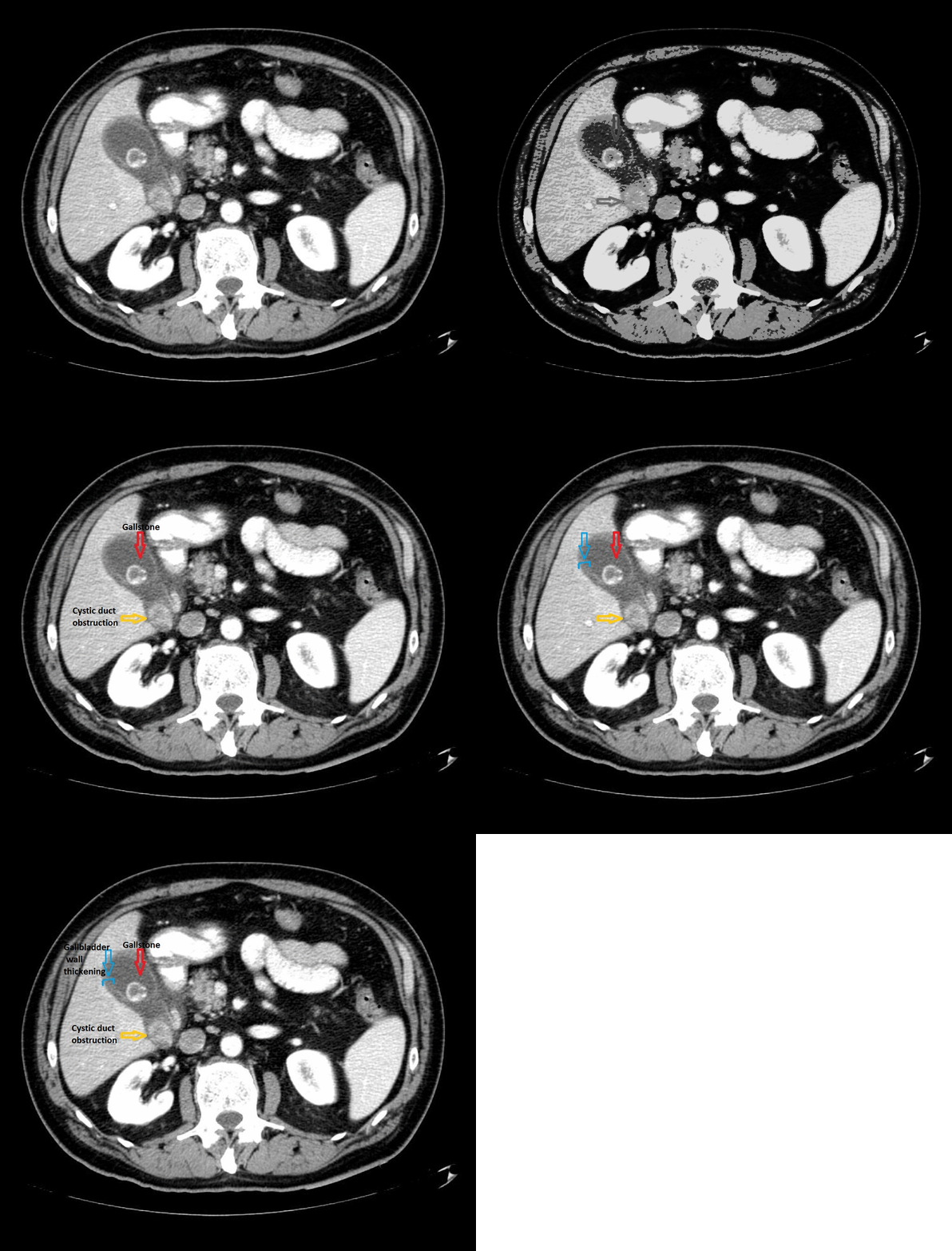
[[File:Acute cholecystitis CT.gif|900px|thumb|center|CT Scan of acute cholecystitis; Red arrow shows gallstones, yellow arrow shows cystic duct obstruction, and blue arrow shows thickening of the gallbladder wall. <small> Case courtesy of Radswiki, Radiopaedia.org, rID: 11161 Source:<ref name="urlAcute cholecystitis | Radiology Reference Article | Radiopaedia.org">{{cite web |url=https://radiopaedia.org/articles/acute-cholecystitis |title=Acute cholecystitis | Radiology Reference Article | Radiopaedia.org |format= |work= |accessdate=}}</ref>]] | |||
==References== | ==References== | ||
| Line 23: | Line 24: | ||
{{WH}} | {{WH}} | ||
{{WS}} | {{WS}} | ||
[[Category: | [[Category: Gastroenterology]] | ||
Latest revision as of 17:07, 28 December 2017
|
Acute cholecystitis Microchapters |
|
Diagnosis |
|---|
|
Treatment |
|
Case Studies |
|
Acute cholecystitis CT scan On the Web |
|
American Roentgen Ray Society Images of Acute cholecystitis CT scan |
|
Risk calculators and risk factors for Acute cholecystitis CT scan |
Editor-In-Chief: C. Michael Gibson, M.S., M.D. [1]; Associate Editor(s)-in-Chief: Furqan M M. M.B.B.S[2]
Overview
CT scan is usually used for the diagnosis of the complications of acute cholecystitis. These complications include emphysematous cholecystitis and gangrenous cholecystitis.
CT scan
- CT scan is usually used for the diagnosis of the complications of acute cholecystitis. These complications include:[1][2][3]
Limitations of CT
- Limitations of CT scan include:[1]
- Helpful in obese patients
- Helpful in patients with gaseous abdominal distention
- Information about other intra-abdominal organs

References
- ↑ 1.0 1.1 Gomes CA, Junior CS, Di Saverio S, Sartelli M, Kelly MD, Gomes CC, Gomes FC, Corrêa LD, Alves CB, Guimarães SF (2017). "Acute calculous cholecystitis: Review of current best practices". World J Gastrointest Surg. 9 (5): 118–126. doi:10.4240/wjgs.v9.i5.118. PMC 5442405. PMID 28603584.
- ↑ Reginelli A, Mandato Y, Solazzo A, Berritto D, Iacobellis F, Grassi R (2012). "Errors in the radiological evaluation of the alimentary tract: part II". Semin. Ultrasound CT MR. 33 (4): 308–17. doi:10.1053/j.sult.2012.01.016. PMID 22824121.
- ↑ "Imaging of Cholecystitis : American Journal of Roentgenology : Vol. 196, No. 4 (AJR)".
- ↑ "Acute cholecystitis | Radiology Reference Article | Radiopaedia.org".