Cavernous angioma pathophysiology

Revision as of 17:40, 3 December 2012 by Charmaine Patel (talk | contribs) (Created page with "__NOTOC__ {{Cavernous angioma}} {{CMG}} Please help WikiDoc by adding more content here. It's easy! Click here to learn about editing. ==Cave...")
(diff) ← Older revision | Latest revision (diff) | Newer revision → (diff)
Jump to navigation Jump to search

Cavernous angioma Microchapters

Home

Patient Information

Overview

Historical Perspective

Pathophysiology

Causes

Differentiating Cavernous angioma from other Diseases

Epidemiology and Demographics

Risk Factors

Natural History, Complications and Prognosis

Diagnosis

History and Symptoms

Physical Examination

CT

MRI

MRA

Other Imaging Findings

Other Diagnostic Studies

Treatment

Medical Therapy

Surgery

Cost-Effectiveness of Therapy

Future or Investigational Therapies

Case Studies

Case #1

Cavernous angioma pathophysiology On the Web

Most recent articles

Most cited articles

Review articles

CME Programs

Powerpoint slides

Images

American Roentgen Ray Society Images of Cavernous angioma pathophysiology

All Images
X-rays
Echo & Ultrasound
CT Images
MRI

Ongoing Trials at Clinical Trials.gov

US National Guidelines Clearinghouse

NICE Guidance

FDA on Cavernous angioma pathophysiology

CDC on Cavernous angioma pathophysiology

Cavernous angioma pathophysiology in the news

Blogs on Cavernous angioma pathophysiology

Directions to Hospitals Treating Cavernous angioma

Risk calculators and risk factors for Cavernous angioma pathophysiology

Editor-In-Chief: C. Michael Gibson, M.S., M.D. [1]

Please help WikiDoc by adding more content here. It's easy! Click here to learn about editing.

Cavernous Angiomas and Venous Angiomas

In up to 30% there is a coincidence of CCM with a venous angioma, also known as a developmental venous anomaly (DVA). These lesions appear either as enhancing linear blood vessels or caput medusae, a radial orientation of small vessels that resemble the hair of Medusa from Greek mythology. These lesions are thought to represent developmental anomalies of normal venous drainage. These lesions should not be removed, as reports of venous infarcts have been reported. When found in association with a CCM that needs resection, great care should be taken not to disrupt the angioma.

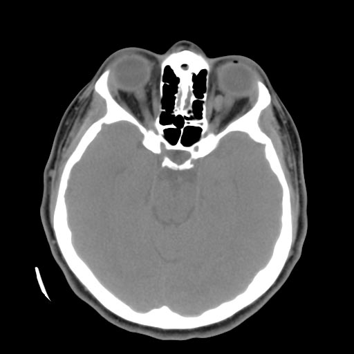
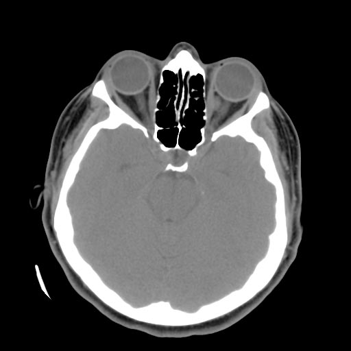
Left orbital cavernous hemangioma
References