Suprarenal veins
| Cardiology Network |
 Discuss Suprarenal veins further in the WikiDoc Cardiology Network |
| Adult Congenital |
|---|
| Biomarkers |
| Cardiac Rehabilitation |
| Congestive Heart Failure |
| CT Angiography |
| Echocardiography |
| Electrophysiology |
| Cardiology General |
| Genetics |
| Health Economics |
| Hypertension |
| Interventional Cardiology |
| MRI |
| Nuclear Cardiology |
| Peripheral Arterial Disease |
| Prevention |
| Public Policy |
| Pulmonary Embolism |
| Stable Angina |
| Valvular Heart Disease |
| Vascular Medicine |
Editor-In-Chief: C. Michael Gibson, M.S., M.D. [1]
Overview
The Suprarenal Veins are two in number:
- the right ends in the inferior vena cava.
- the left ends in the left renal or left inferior phrenic vein.
They receive blood from the adrenal glands and will sometimes form anastomoses with the inferior phrenic veins.
Additional images
-
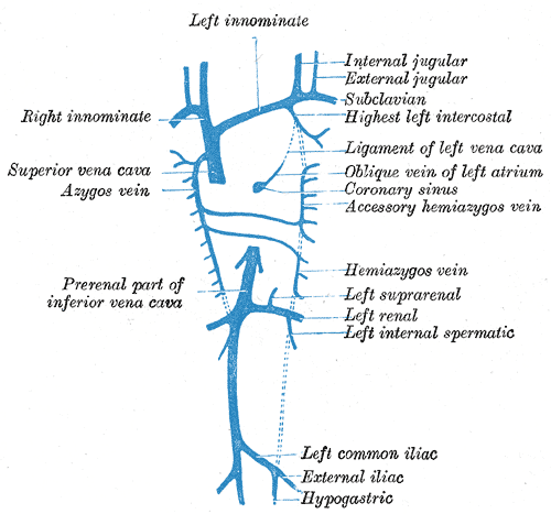
Diagram showing completion of development of the parietal veins.
-
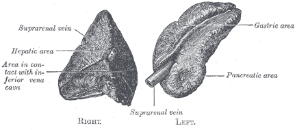
Suprarenal glands viewed from the front.
-
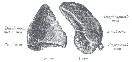
Suprarenal glands viewed from behind.
External links
- Template:SUNYAnatomyLabs - "Posterior Abdominal Wall: Blood Supply to the Suprarenal Glands"
- Template:Dorlands - left
- Template:Dorlands - right


