Medulloblastoma MRI
|
Medulloblastoma Microchapters |
|
Diagnosis |
|---|
|
Treatment |
|
Case studies |
|
Medulloblastoma MRI On the Web |
|
American Roentgen Ray Society Images of Medulloblastoma MRI |
Editor-In-Chief: C. Michael Gibson, M.S., M.D. [1]
Diagnosis
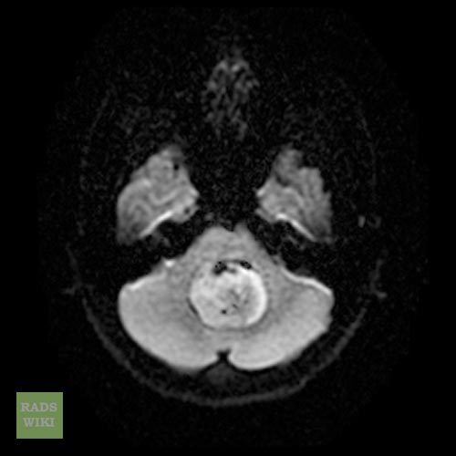
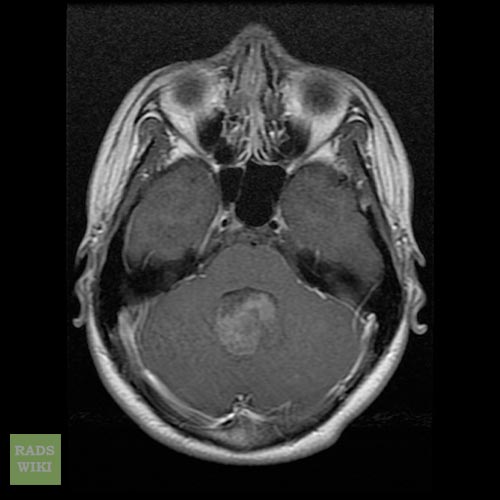
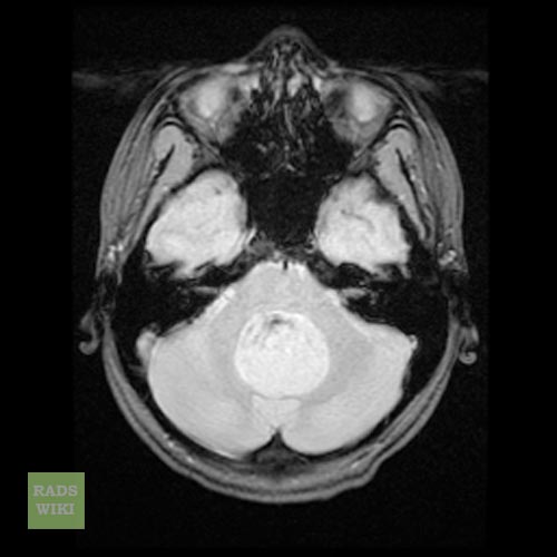
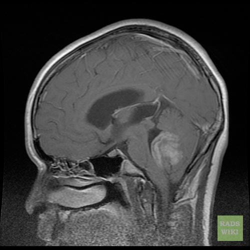
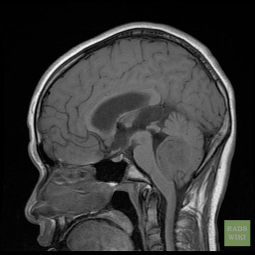
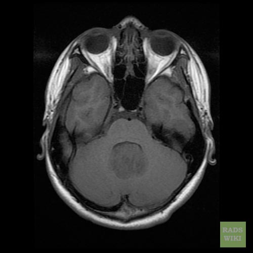
The tumor is distinctive on T1 and T2-weighted MRI with heterogeneous enhancement and typical location adjacent to and extension into the fourth ventricle.
Histologically, the tumor is solid, pink-gray in color, and is well circumscribed. The tumor is very cellular, many mitoses, little cytoplasm, and has the tendency to form clusters and rosettes.
Correct diagnosis of medulloblastoma may require ruling out atypical teratoid rhabdoid tumor (ATRT)[1] and primitive neuroectodermal tumor (PNET).
-
Medulloblastoma
-
Medulloblastoma
-
Medulloblastoma
-
Medulloblastoma
-
Medulloblastoma
-
Medulloblastoma
