Cowden syndrome echocardiography and ultrasound
Editor-In-Chief: C. Michael Gibson, M.S., M.D. [1]; Associate Editor(s)-in-Chief: Vamsikrishna Gunnam M.B.B.S [2]
Overview
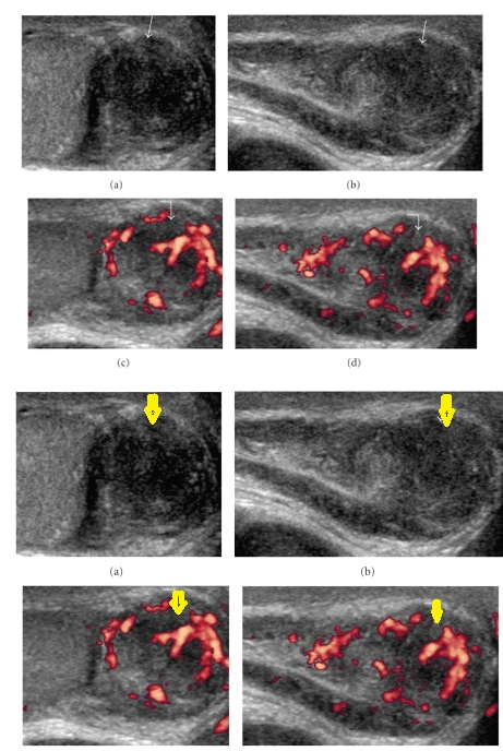
There are ultrasound findings associated with cowden syndrome. Ultrasound may be helpful in the diagnosis of complications of Cowden syndrome, which include testicular swelling, hydrocele, and hyperechoic masses of the testes.
Ultrasound
Ultrasound of the testicles may be helpful in the diagnosis of cowden syndrome. Findings on an ultrasound suggestive of cowden syndrome include:[1]
- Testicular lipomatosis
- Testicular Hamartomas
- Epididymal tumor
- Testicular swelling
- Hydrocele
- Intratesticular hyperechoic masses of varying sizes


References
- ↑ Venkatanarasimha, Nanda; Hilmy, Shakira; Freeman, Simon (2011). "Case 175: Testicular Lipomatosis in Cowden Disease". Radiology. 261 (2): 654–658. doi:10.1148/radiol.11091151. ISSN 0033-8419.
- ↑ "Testicular Hamartomas and Epididymal Tumor in a Cowden Disease: A Case Report".
- ↑ "Testicular Hamartomas and Epididymal Tumor in a Cowden Disease: A Case Report".