Mycosis fungoides pathophysiology
|
Cutaneous T cell lymphoma Microchapters |
Editor-In-Chief: C. Michael Gibson, M.S., M.D. [1]; Associate Editor(s)-in-Chief: Sowminya Arikapudi, M.B,B.S. [2]
Overview
Cutaneous T cell lymphoma arises from T-cell lymphocytes, which are normally involved in the cell mediated immune response. On microscopic histopathological analysis, atypical lymphoid cells, polymorphous inflammatory infiltrate in the dermis, and lymphocytes with cerebroid nuclei are characteristic findings of mycosis fungoides.
Pathophysiology
- Cutaneous T cell lymphoma arises from T-cell lymphocytes, which are normally involved in the cell mediated immune response.
- Sezary syndrome and mycosis fungoides are T-cell lymphomas whose primary manifestation is in the skin
- Mycosis Fungoides is the most common type of cutaneous T cell lymphoma
- Mycosis fungoides is initially an indolent lymphoma but in its later stages can cause peripheral lymphadenopathy and can finally progress to widespread extracutaneous visceral / internal organ involvement
- Sézary's cells are T-cells that have pathological quantities of mucopolysaccharides
- Sézary's disease is sometimes considered a late stage of mycosis fungoides
Gross Pathology
-
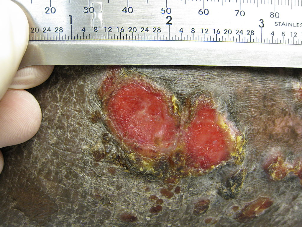
Plaque of mycosis fungoides
-
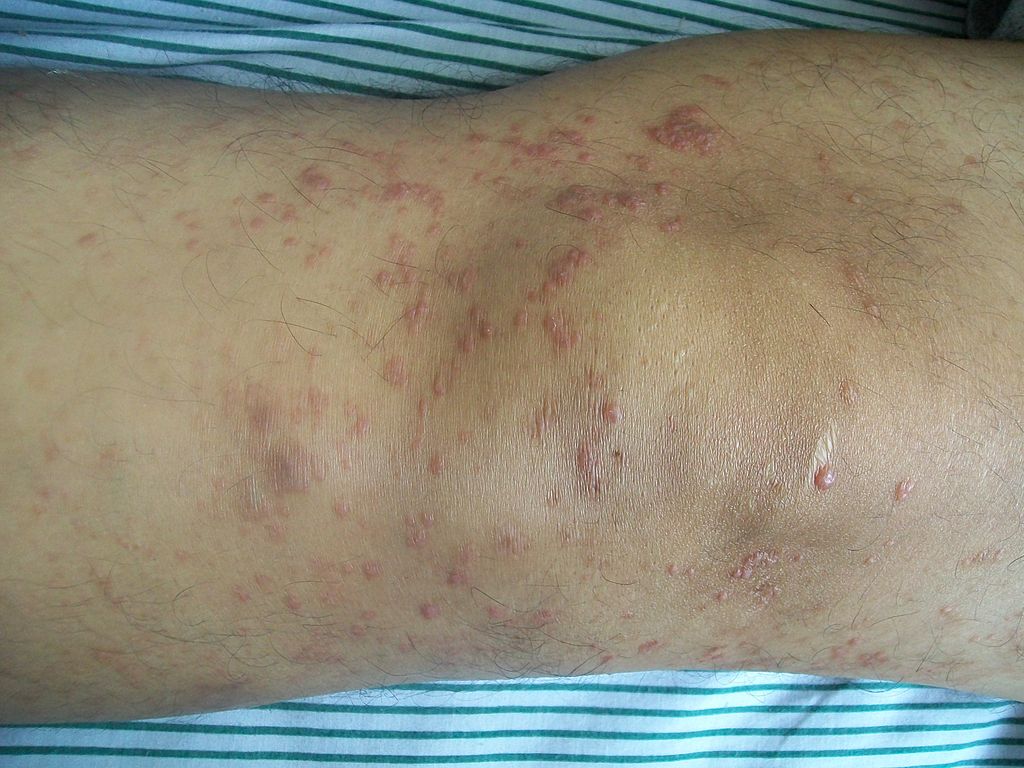
Mycosis fungoides knee


Microscopic Pathology
- Mycosis fungoides has been divided into three stages:
- Premycotic stage
- Mycotic stage
- Tumorous stage
- The premycotic stage
- Non-diagnostic and represented by chronic nonspecific dermatisis associated with psoriasiform changes in epidermis
- The mycotic stage
- Shows a polymorphous inflammatory infiltrate in the dermis that contains small numbers of frankly atypical lymphoid cells
- These cells may line up individually along the epidermal basal layer
- The latter finding if unaccompanied by spongiosis is highly suggestive of mycosis fungoides
- Tumorous stage
- Dense infiltrate of medium sized lymphocytes with cerebroid nuclei, expands the dermis
-
Sézary's disease
-
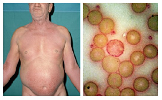
61-year-old man presented in 1972 with unrelenting pruritus of six months’ duration. On the right is his peripheral blood film stained with Periodic Acid-Schiff (PAS) showing a neoplastic T cell (Sézary cell).
-
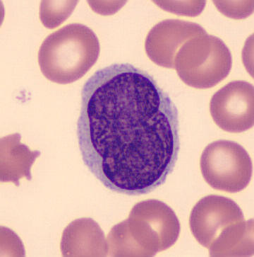
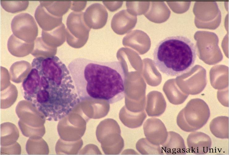
Pleomorphic abnormal T cell with the characteristic cerebriform nuclei (Peripheral blood - MGG stain)
-
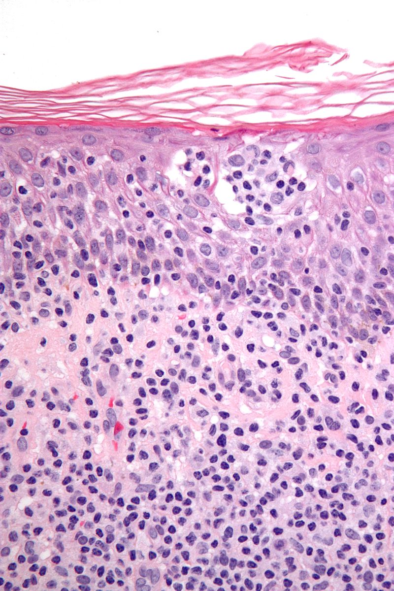
Features: Nests of lymphocytes in the epidermis; "Pautrier microabscesses". Single lymphocytes in epidermis; "lymphocyte exocytosis". Short linear arrays of lymphocytes along the basal layer of the epidermis; "epidermotropism".


