Pheochromocytoma CT
|
Pheochromocytoma Microchapters |
|
Diagnosis |
|---|
|
Treatment |
|
Case Studies |
|
Pheochromocytoma CT On the Web |
|
American Roentgen Ray Society Images of Pheochromocytoma CT |
Editor-In-Chief: C. Michael Gibson, M.S., M.D. [1] Associate Editor(s)-in-Chief: Ahmad Al Maradni, M.D. [2]
Overview
Head, neck, chest and abdomen CT scan may be helpful in the diagnosis of pheochromocytoma.
CT
Computed tomography scan of the head, neck,chest, and abdomen can help localize the tumor.
-
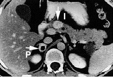
Pheochromocytoma. CT abdomen.
-
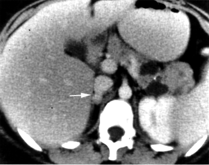
Pheochromocytoma. CT abdomen.


